

The p53 family and programmed cell death
- PMID: 18955976
- PMCID: PMC2657599
- DOI: 10.1038/onc.2008.315
The p53 family and programmed cell death
Abstract
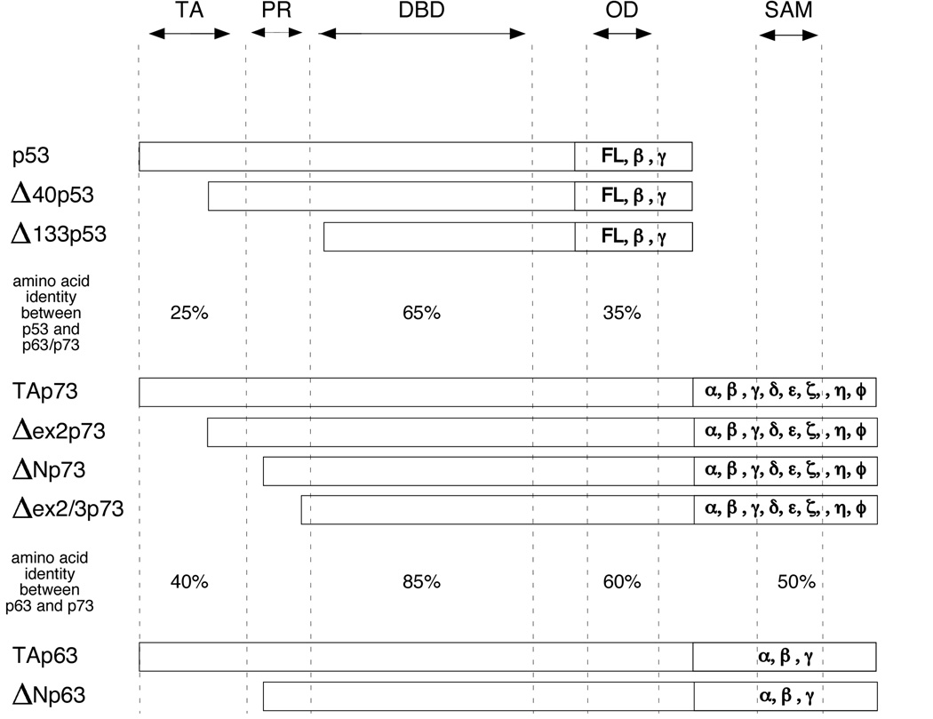
The p53 tumor suppressor continues to hold distinction as the most frequently mutated gene in human cancer. The ability of p53 to induce programmed cell death, or apoptosis, of cells exposed to environmental or oncogenic stress constitutes a major pathway whereby p53 exerts its tumor suppressor function. In the past decade, we have discovered that p53 is not alone in its mission to destroy damaged or aberrantly proliferating cells: it has two homologs, p63 and p73, that in various cellular contexts and stresses contribute to this process. In this review, the mechanisms whereby p53, and in some cases p63 and p73, induce apoptosis are discussed. Other reviews have focused more extensively on the contribution of individual p53-regulated genes to apoptosis induction by this protein, whereas in this review, we focus more on those factors that mediate the decision between growth arrest and apoptosis by p53, p63 and p73, and on the post-translational modifications and protein-protein interactions that influence this decision.
Figures

References
-
- Agami R, Blandino G, Oren M, Shaul Y. Interaction of c-Abl and p73alpha and their collaboration to induce apoptosis. Nature. 1999;399:809–813. - PubMed
-
- Bargonetti J, Manfredi JJ. Multiple roles of the tumor suppressor p53. Curr Opin Oncol. 2002;14:86–91. - PubMed
-
- Barlev NA, Liu L, Chehab NH, Mansfield K, Harris KG, Halazonetis TD, Berger SL. Mol Cell. 2001;8:1243–1254. - PubMed
-
- Basu S, Totty NF, Irwin MS, Sudol M, Downward J. Akt phosphorylates the Yes-associated protein, YAP, to induce interaction with 14-3-3 and attenuation of p73-mediated apoptosis. Mol Cell. 2003;11:11–23. - PubMed
Publication types
MeSH terms
Substances
Grants and funding
LinkOut - more resources
Full Text Sources
Other Literature Sources
Research Materials
Miscellaneous
