Review
. 2008 Nov;28(4):360-9.
doi: 10.1055/s-0028-1091980. Epub 2008 Oct 27.

Molecular mechanisms of lipotoxicity in nonalcoholic fatty liver disease

Affiliations
Review

Molecular mechanisms of lipotoxicity in nonalcoholic fatty liver disease

Harmeet Malhi et al. Semin Liver Dis. 2008 Nov.

Abstract

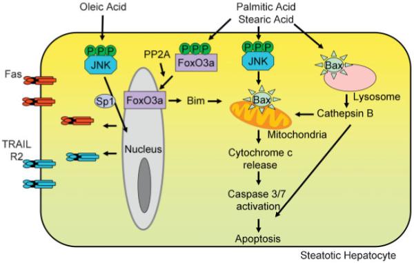
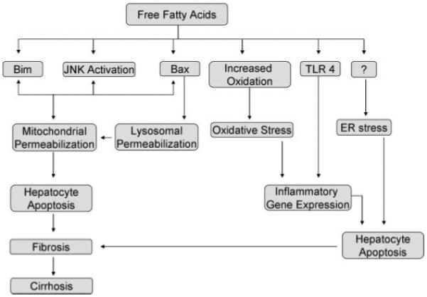
Nonalcoholic fatty liver disease (NAFLD) is characterized by insulin resistance, which results in elevated serum concentration of free fatty acids (FFAs). Circulating FFAs provide the substrate for triacylglycerol formation in the liver, and may also be directly cytotoxic. Hepatocyte apoptosis is a key histologic feature of NAFLD, and correlates with progressive inflammation and fibrosis. The molecular pathways leading to hepatocyte apoptosis are not fully defined; however, recent studies suggest that FFA-induced apoptosis contributes to the pathogenesis of nonalcoholic steatohepatitis. FFAs directly engage the core apoptotic machinery by activating the proapoptotic protein Bax, in a c-jun N-terminal kinase-dependent manner. FFAs also activate the lysosomal pathway of cell death and regulate death receptor gene expression. The role of ER stress and oxidative stress in the pathogenesis of nonalcoholic steatohepatitis has also been described. Understanding the molecular mediators of liver injury should promote development of mechanism-based therapeutic interventions.


Figures

Figure 1
Figure 1
Free fatty acids (FFAs) induce hepatocyte apoptosis. FFAs can modulate both the extrinsic and the intrinsic pathways of hepatocyte apoptosis. The saturated fatty acids, palmitic acid and stearic acid, lead to c-jun N-terminal kinase (JNK) dependent activation of the proapoptotic protein Bax, which then leads to mitochondrial permeabilization with release of cytochrome c, activation of effector caspases, and apoptosis. Palmitic acid can also activate the lysosomal pathway of apoptosis, via Bax activation and Bax-dependent lysosomal permeabilization. Furthermore, palmitic acid and stearic acid activate protein phosphatase 2A (PP2A) leading to activation of FoxO3a, and transcriptional activation of the proapoptotic protein Bim. The monounsaturated fatty acid, oleic acid, which is minimally toxic per se, imparts sensitivity to the death receptor mediated extrinsic pathway of apoptosis. Fas and TRAIL-R2 expression is induced by oleic acid treatment. TRAIL-R2 expression is under the transcriptional control of JNK in oleic acid treated cells. This sensitizes fatty hepatocytes to circulating Fas or TRAIL.
Figure 2
Figure 2
Free fatty acids (FFAs) and their molecular targets. FFA toxicity occurs at multiple molecular levels. Palmitic acid and stearic acid induce Bim expression, under transcriptional control of FoxO3a. Free fatty acids can activate c-jun N-terminal kinase (JNK), and the magnitude of induction correlates with their toxicity. JNK activation leads to Bax-induced mitochondrial permeabilization. Bax can also lead to lysosomal permeabilization and apoptosis. Increased oxidation of fatty acids promotes the formation of reactive oxygen species, resulting in antioxidant depletion and oxidative stress. Toll-like receptor 4 is activated by palmitic acid and oleic acid, thus regulating inflammatory gene expression. Endoplasmic reticulum stress is also a feature of human nonalcoholic fatty liver disease and can be induced by saturated fatty acids, palmitic acid and stearic acid; however, the mechanisms are unclear.

References

    1. Unger RH. Minireview: weapons of lean body mass destruction—the role of ectopic lipids in the metabolic syndrome. Endocrinology. 2003;144(12):5159–5165. - PubMed
    1. Hotamisligil GS. Inflammation and metabolic disorders. Nature. 2006;444(7121):860–867. - PubMed
    1. Gregor MF, Hotamisligil GS. Thematic review series: adipocyte biology. adipocyte stress: the endoplasmic reticulum and metabolic disease. J Lipid Res. 2007;48(9):1905–1914. - PubMed
    1. Jaworski K, Sarkadi-Nagy E, Duncan RE, Ahmadian M, Sul HS. Regulation of triglyceride metabolism: IV. Hormonal regulation of lipolysis in adipose tissue. Am J Physiol Gastrointest Liver Physiol. 2007;293(1):G1–G4. - PMC - PubMed
    1. Baranova A, Gowder SJ, Schlauch K, et al. Gene expression of leptin, resistin, and adiponectin in the white adipose tissue of obese patients with non-alcoholic fatty liver disease and insulin resistance. Obes Surg. 2006;16(9):1118–1125. - PubMed

Publication types

MeSH terms

Substances