. 2008;52(7):985-91.
doi: 10.1387/ijdb.072477ea.

Isolation and expression analysis of foxj1 and foxj1.2 in zebrafish embryos

Affiliations

Isolation and expression analysis of foxj1 and foxj1.2 in zebrafish embryos

Emil Aamar et al. Int J Dev Biol. 2008.

Abstract

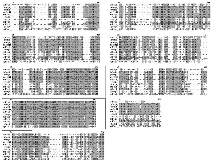
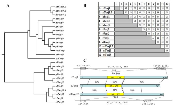
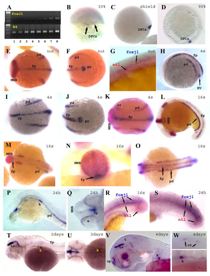
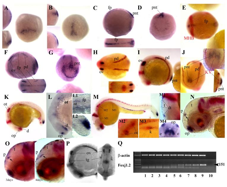
In this report, we present the isolation and identification of a zebrafish homolog of the winged helix\forkhead transcription factor Foxj1. Foxj1 was identified in other species but not in zebrafish. Foxj1 was shown in mice to be required in ciliogenesis and left-right asymmetry establishment. Here we present a spatio-temporal expression pattern of zebrafish foxj1, showing that this gene is expressed in dorsal forerunner cells, Kupffers vesicle, the floor plate, pronephric ducts and kidney. This expression pattern is overlapping but different from that of the foxj1.2, the closest related gene in zebrafish. Foxj1 in zebrafish appears to have similar functions as those reported in other species connected to ciliogenesis and left-right asymmetry.


Figures

Fig. 1
Fig. 1. Molecular analysis of Foxj1
Amino acid alignment of predicted zebrafish Foxj1 protein with Foxj1 in other species, using the http://molbio.info.nih.gov/molbio/gcglite/clustal17.html online program. Consensus amino acids are shaded. The Winged helix domain is boxed. Zebrafish Foxj1 (ABW82682) aligns at a level of 99% to the hypothetical sequence (AAI24229). h, Homo sapiens (AAB09039); p, Pan troglodytes (XP_511694); ma, Macaca mulatta (XP_001104114); m, Mus musculus (AAH82543); r, Rattus norvegicus (NP_446284); xl, Xenopus laevis (AAH77846); xt, Xenopus tropicalis (CAJ82756); cf, Canis familiaris (XP_533124); g, Gallus gallus (XP_001233327); and sp, Strongylocentrotus purpuratus (NP_001073013).
Fig. 2
Fig. 2. Comparison of Foxj protein sequences
A) Phylogenic tree of Foxj1/1.2/2/ and 3 proteins, given by http://align.genome.jp/ CLUSTALW. B) “PairWise” Comparisons Scores (percentage). C) Schematic drawing of identities between zebrafish Foxj1 and Foxj1.2 and mouse Foxj1. The forkhead box is in yellow (FH Box), and chromosomal locations of zebrafish genes are indicated; proteins are encoded by two exons. The accession numbers used in these comparisons are as follows (the numbers for Foxj1 are as in Figure 1): Danio rerio (zebrafish, zf) Foxj1.2 (NP_001008648.1); Homo sapiens (h) Foxj2 (NP_060886) and Foxj3 (Q9UPW0); Mus musculus (m), Foxj2 (NP_068699.1), and Foxj3 (Q8BUR3); Rattus norvegicus (r), Foxj2 (XP_578387.2), and Foxj3 (NP_001101441); Xenopus tropicalis (xt), Foxj1.2 (Q66IG8), Foxj2 (Q28EM1), and Foxj3 (NP_001103516); Xenopus laevis (xl), Foxj1.2 (Q32NH9), and Foxj2 (NP_001087521.1), Gallus gallus (g), Foxj2 (XP_416484); Canis familiaris (cf), Foxj3.1 (XP_532540).
Fig. 3
Fig. 3. Expression pattern of foxj1
A)RT-PCR expression analysis of zebrafish foxj1 and histone 4 (H4) as control was performed for different stages (1-8: unfertilized eggs, 100-200 cells, high-dome, 40-50% epiboly, 80-90%, bud, 6-somites, and 24h embryos, respectively). B-W) Spatio-temporal expression pattern of zebrafish foxj1. Whole-mount in situ hybridization with zebrafish foxj1 probe alone (B-D, H-J, P-Q, T-W), or combined with either pax2.1 (red: E-F, K-O) or ntl (red: G, R-S). Stages are indicated at top right, with “s” referring to somite number, and hpf referring to hours post-fertilization. Views are as follows: (B) dorsal, (C, D) lateral with dorsal to the right, (E, I, K, M, O, U) dorsal with anterior to the left, (F, J) posterior with dorsal to the left, (Q) anterior with dorsal to the left, (G, H, L, P, R, S, T, V, W) lateral with anterior to the left, and (N) is posterior with dorsal up. DFCs, dorsal forerunner cells; e, eye; fp, floor plate; k, kidney; KV, Kupffer’s vesicle; MHB, mid-hindbrain boundary; op, olfactory pits; ot; otolith; ov, otic vesicle; pd, pronephric duct; t, tectum; tv, tectal ventricle.
Fig. 4
Fig. 4. Zebrafish foxj1.2 expression
A-P) Spatio-temporal expression pattern of foxj1.2. Whole-mount in situ hybridization with foxj1.2 probe alone (A-D, F-G, L, M4), or combined with pax2.1 and charon (both red: E, H-K, M-P). Shield-60% epiboly (A), 80% epiboly (B), bud (C-E), 4 somites (F-G), 7 somites (H-J), 15 somites (K, P), 16 somites (L), 1 day (M), 2 days (N), 3 and 4 days (P left and right respectively). Views are as follows: dorsal (A, B); dorsal with anterior to the left (C bottom, E, F bottom, H and H bottom), dorsal with anterior up (P right), lateral with dorsal to the right, (A left bottom, B left bottom), lateral with anterior to left (C top, F, I, K-O except for M4, P left), posterior with dorsal up (D, G, J), anterior ventral with dorsal up (M4), animal view (A left top, B left top), ventral with posterior to the right (J right bottom). Q) RT-PCR expression analysis of foxj1.2 and β-actin as control was performed for different stages (1-8: unfertilized eggs, 100-200 cells, high-dome, 40-50% epiboly, 80-90%, bud, 13-somites, 24hpf and 3days old embryos respectively, and -RT in lane 10). d, diencephalon; e, eye; fp, floor plate; k, kidney; KV, Kupffer’s vesicle; MHB, mid-hindbrain boundary; nt, notochord; op, olfactory pit; ot; otolith; ov, otic vesicle; pd, pronephric duct; pnt, posterior neural tube; t, tectum; tv, tectal ventricle.

References

    1. Avraham KB, Fletcher C, Overdier DG, Clevidence DE, Lai E, Costa RH, Jenkins NA, Copeland NG. Murine chromosomal location of eight members of the hepatocyte nuclear factor 3/fork head winged helix family of transcription factors. Genomics. 1995;25:388–93. - PubMed
    1. Blatt EN, Yan XH, Wuerffel MK, Hamilos DL, Brody SL. Forkhead transcription factor HFH-4 expression is temporally related to ciliogenesis. Am J Respir Cell Mol Biol. 1999;21:168–76. - PubMed
    1. Brody SL, Hackett BP, White RA. Structural characterization of the mouse Hfh4 gene, a developmentally regulated forkhead family member. Genomics. 1997;45:509–18. - PubMed
    1. Brody SL, Yan XH, Wuerffel MK, Song SK, Shapiro SD. Ciliogenesis and left-right axis defects in forkhead factor HFH-4-null mice. Am J Respir Cell Mol Biol. 2000;23:45–51. - PubMed
    1. Chen J, Knowles HJ, Hebert JL, Hackett BP. Mutation of the mouse hepatocyte nuclear factor/forkhead homologue 4 gene results in an absence of cilia and random left-right asymmetry. J Clin Invest. 1998;102:1077–82. - PMC - PubMed

Publication types

Substances