. 2008 Nov 25;47(47):12290-8.
doi: 10.1021/bi800555z.

NMR solution structure of the neurotrypsin Kringle domain

Affiliations

NMR solution structure of the neurotrypsin Kringle domain

Olga A Ozhogina et al. Biochemistry. .

Abstract

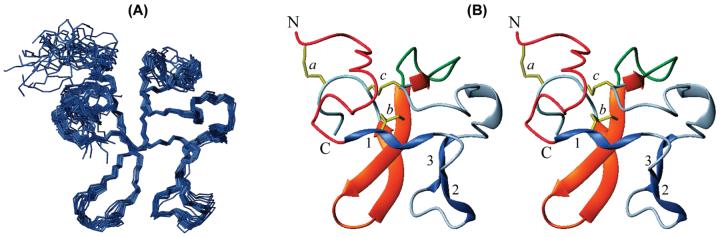
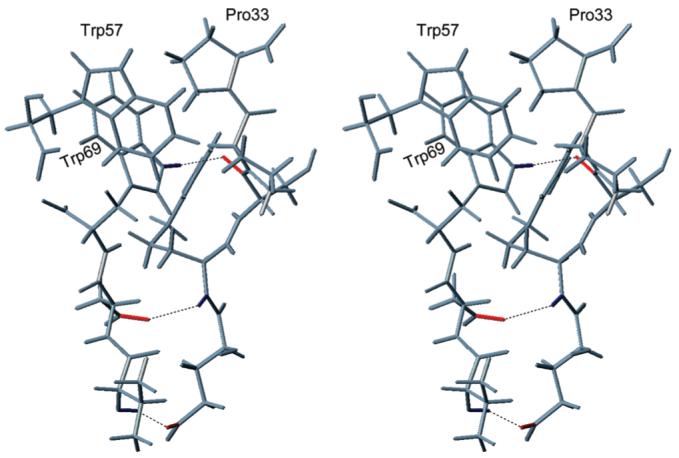
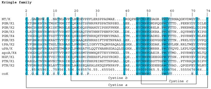
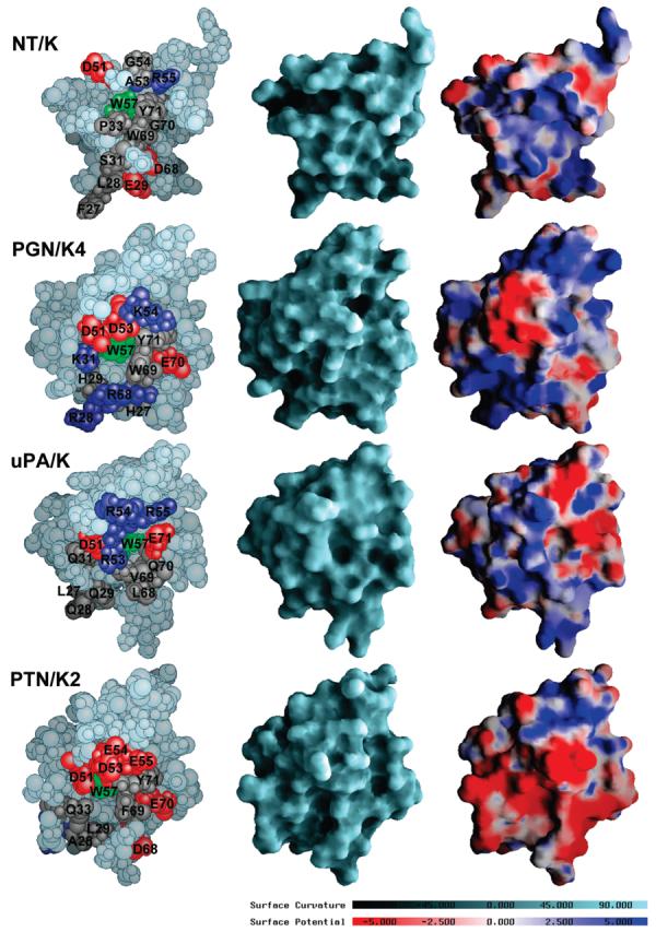
Neurotrypsin is a multidomain protein that serves as a brain-specific serine protease. Here we report the NMR structure of its kringle domain, NT/K. The data analysis was performed with the BACUS (Bayesian analysis of coupled unassigned spins) algorithm. This study presents the first application of BACUS to the structure determination of a 13C unenriched protein for which no prior experimental 3D structure was available. NT/K adopts the kringle fold, consisting of an antiparallel beta-sheet bridged by an overlapping pair of disulfides. The structure reveals the presence of a surface-exposed left-handed polyproline II helix that is closely packed to the core beta-structure. This feature distinguishes NT/K from other members of the kringle fold and points toward a novel functional role for a kringle domain. Functional divergence among kringle domains is discussed on the basis of their surface and electrostatic characteristics.


Figures

FIGURE 1
FIGURE 1
(A) Superposition of the backbone atoms (N, Cα, C′) of residues 8–74 in the ensemble of 30 NT/K structures. (B) Stereoview of ribbon diagram showing the secondary structure elements in NT/K. The disulfides (yellow) are labeled a, b, and c; the PPII-helices (blue) are designated 1 (residues 17–20), 2 (residues 24–26), and 3 (residues 31–33); β-strands (residues 57–62, 66–71) and a single bridge (residues 48–57) are shown in orange; green loop corresponds to residues 47–56; N- and C-termini are in red and light blue, respectively.
FIGURE 2
FIGURE 2
Stereoview of fragment of NT/K containing the hydrogen bonds 30→68, 67→29, and 69→31 which accommodate Pro33 between the side chains of Trp57 and Trp69.
FIGURE 3
FIGURE 3
Multiple sequence alignments of the kringle family: NT/K (this work), PGN/K1-K3 (code 1ki0), PGN/K4 (code 1pk4), PGN/K5 (code 5hpg), tPA/K2 (code 1tpk), uPA/K (code 2fd6), apoA/K4 (code 1i71), HGF/K1 (code 1bht), PTN/K1 (code 1nl1), and PTN/K2 (code 2hpp). The numbering in the top row refers to the sequential numbering of NT/K. The columns are shaded according to the conservation scores given in the second row from the bottom (e.g., 6 corresponds to a rounded score of 0.6, 7 to a rounded score of 0.7, etc., and 0 to the highest score of 1.0). Positions in which the conservation score is lower than 0.5 are indicated with a dot.
FIGURE 4
FIGURE 4
Comparison of molecular surface features of NT/K (this work), PGN/K4 (code 1pk4) (33), uPA/K (code 2fd6) (35) and PTN/K2 (code 2hpp) (34). The structures were superimposed using cystines b and c and depicted in the 180° orientation relative to the ribbon representation of the folding of NT/K shown in Figure 1B. The views on the left are space-filling atomic models. Residues discussed in the text have been highlighted: positively charged residues (Arg and Lys) are in dark blue, negatively charged residues (Asp and Glu) are in red, neutral residues are in gray, and the conserved residue W57 is in green. The views in the center are representations of the accessible molecular surfaces. The surfaces are color coded according to curvature with convex areas in aquamarine, the planar regions in light gray, and concave areas in dark gray. The views on the right are depictions of the electrostatic surface potentials. The potentials were calculated at 150 mM NaCl using DelPhi (30), interpolated onto the molecular surfaces, and visualized with GRASP (30), using a scale from −5 kBT/e (dark red) to +5 kBT/e (dark blue), where kB is the Boltzmann constant, T is ambient temperature, and e is the unit charge.

References

    1. Murzin AG, Brenner SE, Hubbard TJP, Chothia C. SCOP: a structural classification of proteins database for the investigation of sequences and structures. J. Mol. Biol. 1995;247:536–540. - PubMed
    1. McLean JW, Tomlinson JE, Kuang WJ, Eaton DL, Chen EY, Fless GM, Scanu AM, Lawn RM. cDNA sequence of human apolipoprotein A is homologous to plasminogen. Nature. 1987;330:132–137. - PubMed
    1. Waisman DM, editor. Plasminogen: Structure, Activation, and Regulation. Springer-Verlag; New York: 2003.
    1. Blasi F, Carmeliet P. uPAR: A versatile signalling orchestrator. Nat. Rev. Mol. Cell Biol. 2002;3:932–943. - PubMed
    1. Bock GR, Goode JA, editors. Ciba Foundation Symposium Series. Wiley; New York: 1998. Plasminogen-Related Growth Factors.

Publication types