. 2008 Dec;107(5):1436-47.
doi: 10.1111/j.1471-4159.2008.05708.x. Epub 2008 Oct 24.

Rit signaling contributes to interferon-gamma-induced dendritic retraction via p38 mitogen-activated protein kinase activation

Affiliations

Rit signaling contributes to interferon-gamma-induced dendritic retraction via p38 mitogen-activated protein kinase activation

Douglas A Andres et al. J Neurochem. 2008 Dec.

Abstract

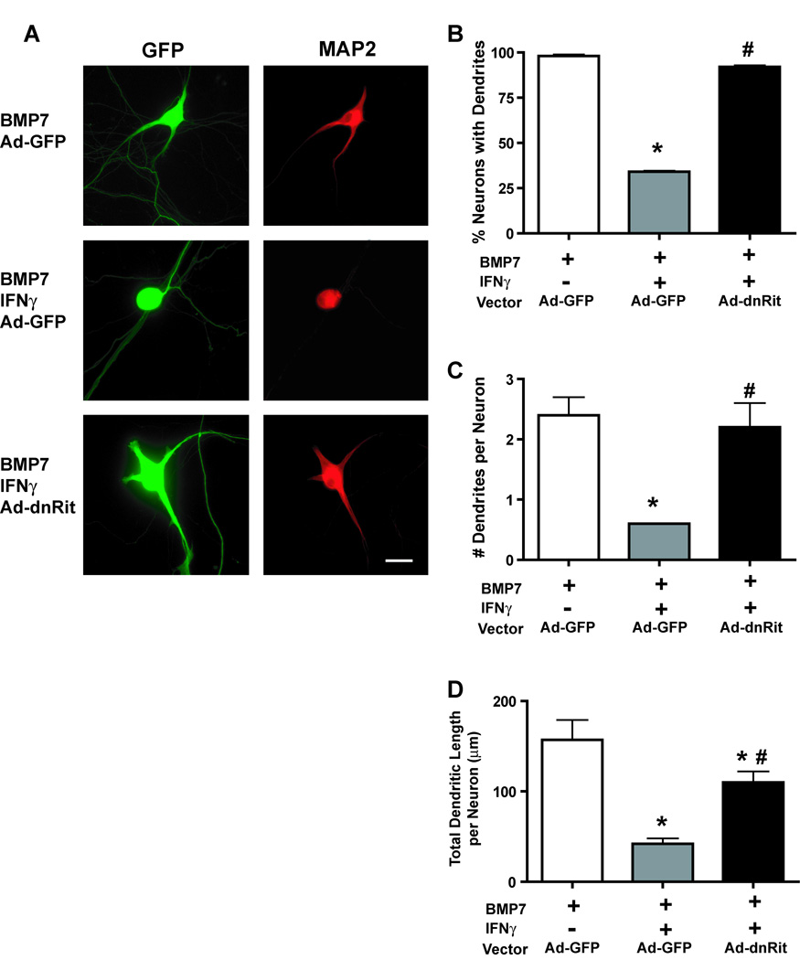
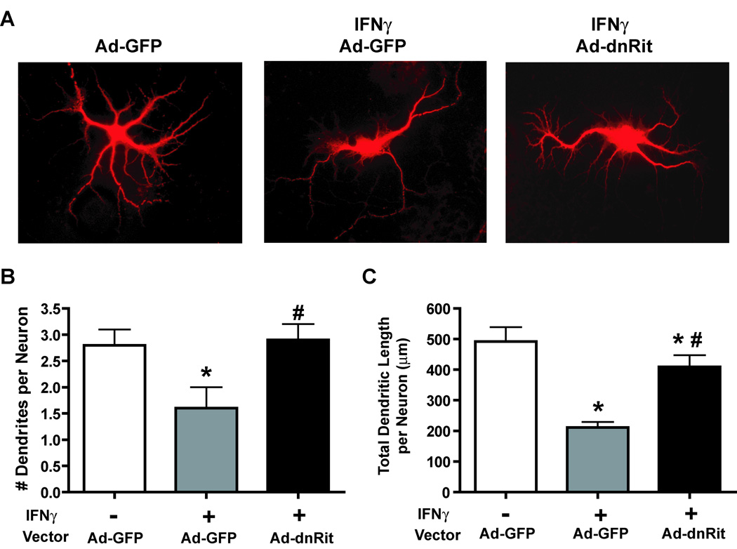
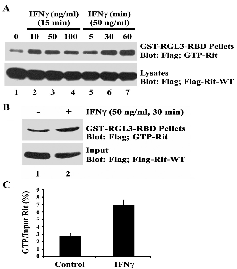
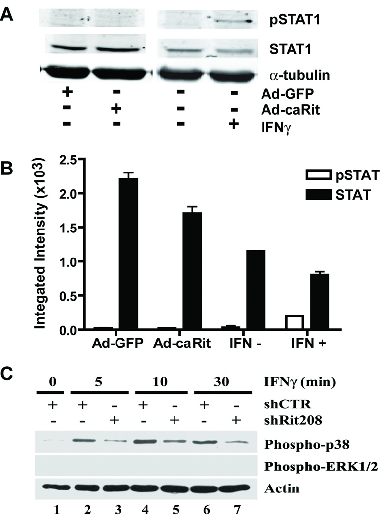
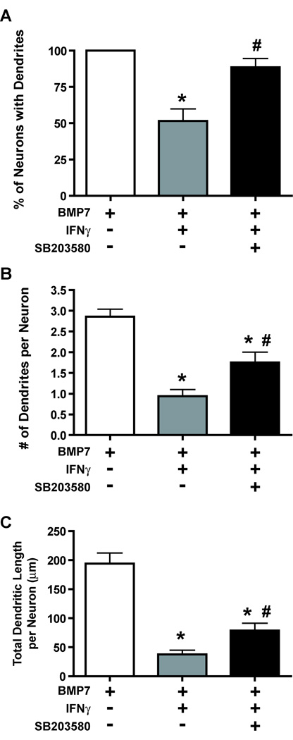
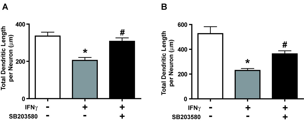
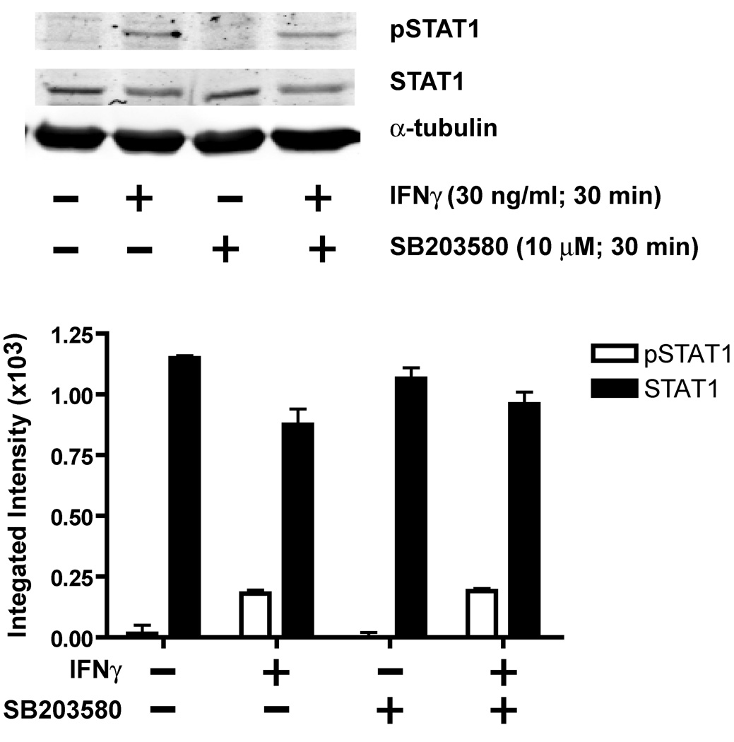
The proinflammatory cytokine interferon-gamma (IFNgamma) alters neuronal connectivity via selective regressive effects on dendrites but the signaling pathways that mediate this effect are poorly understood. We recently demonstrated that signaling by Rit, a member of the Ras family of GTPases, modulates dendritic growth in primary cultures of sympathetic and hippocampal neurons. In this study, we investigated a role for Rit signaling in IFNgamma-induced dendritic retraction. Expression of a dominant negative Rit mutant inhibited IFNgamma-induced dendritic retraction in cultured embryonic rat sympathetic and hippocampal neurons. In pheochromacytoma cells and hippocampal neurons, IFNgamma caused rapid Rit activation as indicated by increased GTP binding to Rit. Silencing of Rit by RNA interference suppressed IFNgamma-elicited activation of p38 MAPK in pheochromacytoma cells, and pharmacological inhibition of p38 MAPK significantly attenuated the dendrite-inhibiting effects of IFNgamma in cultured sympathetic and hippocampal neurons without altering signal transducer and activator of transcription 1 activation. These observations identify Rit as a downstream target of IFNgamma and suggest that a novel IFNgamma-Rit-p38 signaling pathway contributes to dendritic retraction and may, therefore, represent a potential therapeutic target in diseases with a significant neuroinflammatory component.


Figures

Figure 1
Figure 1. Inhibiting Rit activation blocks IFNγ-induced dendrite retraction in cultured sympathetic neurons
(A) Fluorescence micrographs of sympathetic neurons immunostained for GFP and MAP2. Sympathetic neurons were initially treated with BMP7 (50 ng/ml) for 5 days to induce dendritic growth, and then infected with adenovirus expressing GFP alone (Ad-GFP) or co-expressing GFP and dominant-negative RitS35N (Ad-dnRit). IFNγ (30 ng/ml) was added to a subset of cultures immediately following infection. Cultures were fixed 3 days after infection and immunostained for GFP and MAP2, a cytoskeletal protein localized to the somatodendritic compartment. Sympathetic neurons infected with Ad-GFP and exposed to BMP7 alone extended several MAP2 immunopositive processes. Addition of IFNγ caused dendritic retraction in neurons expressing Ad-GFP. Expression of dnRit significantly attenuated dendrite retraction in cultures exposed to IFNγ. Dendritic growth was quantified in neuronal cells dual labeled for GFP and MAP2 with respect to the percentage of neurons with dendrites (B), the number of dendrites per neuron (C) and the total length of the dendritic arbor per neuron (D). Data are presented as the mean ± SEM (n > 80 neurons per experimental group). * Significantly different from Ad-GFP neurons exposed to BMP7 at p < 0.05; # significantly different from Ad-GFP neurons treated with BMP7 and IFNγ at p < 0.05.
Figure 2
Figure 2. Inhibiting Rit activation blocks IFNγ-induced dendrite retraction in cultured hippocampal neurons
(A) Fluorescence micrographs of GFP-positive hippocampal neurons immunostained for MAP2 (MAP2 immunoreactivity is shown). Hippocampal neurons were infected with adenovirus expressing GFP alone (Ad-GFP) or co-expressing GFP and dominant-negative RitS35N (Ad-dnRit) 4 days after plating. IFNγ (30 ng/ml) was added to a subset of cultures immediately following infection. Cultures were fixed 3 days after infection and immunostained for both GFP (green) and MAP2 (red). IFNγ caused dendritic retraction in hippocampal neurons infected with Ad-GFP. Expression of dnRit significantly inhibited the dendrite retracting activity of IFNγ. Dendritic growth was quantified in neuronal cells immunopositive for both GFP and MAP2 with respect to the number of dendrites per neuron (B) and the total length of the dendritic arbor per neuron (C). Data are presented as the mean ± SEM (n > 80 neurons per experimental group). *Significantly different from Ad-GFP neurons maintained in the absence of IFNγ at p < 0.05; # significantly different from Ad-GFP neurons treated with IFNγ at p < 0.05.
Figure 3
Figure 3. IFNγ activates Rit in PC6 cells and in cultured hippocampal neurons
GST pull-down assays using GST-RGL3-RBD agarose were performed with lysates obtained from PC6 cells (A) or hippocampal neurons (B) expressing 3xFlag-Rit-WT. The levels of GTP-bound Rit were determined by immunoblot analysis using anti-Flag monoclonal antibody. (A) PC6 cells were serum starved for 5 h to reduce basal Rit-GTP levels prior to stimulation with increasing concentrations of IFNγ for 15 min or with IFNγ at 50 ng/ml for 0, 5, 30 or 60 min. Note that Rit-GTP levels increase following exposure to IFNγ in a time- and concentration-dependent manner. (B) Hippocampal neurons were transfected with 3xFlag-Rit-WT at the time of plating, maintained in serum-free Neurobasal medium supplemented with B27 for 72 h then either left untreated or exposed to IFNγ at 50 ng/ml for 30 min. Rit is activated in hippocampal neurons treated with IFNγ as illustrated in a representative blot (B) and confirmed by densitometry (C) in which values obtained for Rit-GTP are normalized to total Flag-Rit-WT values (data presented as the mean ± SEM, n = 2 per experimental condition). In experiments using either PC6 cells or hippocampal neurons, similar results were obtained in two separate experiments performed using two different sets of cultures.
Figure 4
Figure 4. Rit activates p38, but neither ERK1/2 nor STAT signaling
(A and B) Hippocampal neurons were infected with adenovirus expressing GFP alone (Ad-GFP) or co-expressing GFP and constitutively active RitQ79L (Ad-caRit) 4 days after plating. Cell lysates were collected 16 h post-infection and analyzed by immunoblotting using mAb that specifically recognize phosphorylated Ser727 on STAT1 (pSTAT1) or polyclonal antibodies that react with both phosphorylated and nonphosphorylated STAT1 (STAT1). A representative Western blot (A) and corresponding densitometric analysis (B) of pSTAT1 and total STAT1 indicate that treatment with IFNγ but not expression of caRit activates STAT1 as indicated by increased levels of pSTAT1. Data are expressed as mean ± SEM (n=3 blots obtained in 3 separate experiments performed using cultures from 3 separate dissections). (C) IFNγ activates p38 but not ERK1/2 in PC6 cells. PC6 cells were transfected with either shCTR or shRit208 RNAi vectors and subjected to G418 selection for 48 h. Cells were then serum starved (serum-free DMEM, 5 h) prior to stimulation with IFNγ (50 ng/ml). Whole cell lysates were prepared and levels of activated p38 and ERK1/2 determined by phosphospecific immunoblotting. A representative Western blot for activated p38 and ERK1/2 MAP kinase levels is shown. Anti-actin immunoblotting was used to confirm equivalent protein loading. Note that IFNγ fails to stimulate ERK MAP kinase signaling in PC6 cells and that Rit silencing inhibits IFNγ-mediated p38 activation.
Figure 5
Figure 5. Pharmacological inhibition of p38 kinase attenuates IFNγ-induced dendrite retraction in cultured sympathetic neurons
Sympathetic neurons were pretreated with BMP7 (50 ng/ml) for 5 days to induce dendritic growth. Subsequently, they were divided into 3 groups that were treated for an additional 3 days with: 1) BMP7 alone; 2) BMP7 and IFNγ (30 ng/ml) or 3) BMP7 and IFNγ in the presence of the p38 kinase inhibitor SB203580 (10 µM). Dendritic growth was quantified in neuronal cells immunopositive for MAP2 with respect to the percentage of neurons with dendrites (A), the number of dendrites per neuron (B), and the total dendritic length per neuron (C). Data are expressed as the mean ± SEM (n ≥ 80 neurons per experimental group). *Significantly different from BMP7 alone at p < 0.05; # significantly different from BMP7 and IFNγ at p < 0.05.
Figure 6
Figure 6. IFNγ-induced dendrite retraction in cultured hippocampal neurons is significantly attenuated by a p38 kinase inhibitor
Hippocampal neurons were treated for 3 days with IFNγ (30 ng/ml) in the absence or presence of the p38 kinase inhibitor SB203580 (10 µM). Treatments were initiated on either day 4 in vitro to block initial stages of dendritic growth (A) or on day 7 in vitro to cause retraction of existing dendrites (B). Cultures were fixed and immunostained for the dendritic marker MAP2 and dendritic growth quantified with respect to the total dendritic length per neuron. Data are expressed as the mean ± SEM (n ≥ 80 neurons per experimental group). * Significantly different from BMP7 alone at p < 0.05; # significantly different from BMP7 and IFNγ at p < 0.05.
Figure 7
Figure 7. IFNγ-mediated STAT activation does not require p38 signaling
On day 7 in vitro, hippocampal neurons were treated with IFNγ (30 ng/ml) for 30 min in the absence or presence of the p38 MAP kinase inhibitor, SB203580 (10 µM). Cell lysates were analyzed by immunoblotting using phosphospecific antibodies for STAT1. A representative Western blot (top panel) and corresponding densitometric analysis (bottom panel) of STAT1 phosphorylated on Ser727 (pSTAT) and total (phosphorylated and non-phosphorylated) STAT1 indicate that SB203580 does not block IFNγ activation of STAT1. Data are expressed as mean ± SEM (n=3 blots obtained in 3 separate experiments performed using cultures from 3 separate dissections).

References

    1. Arendt T, Gartner U, Seeger G, Barmashenko G, Palm K, Mittmann T, Yan L, Hummeke M, Behrbohm J, Bruckner MK, Holzer M, Wahle P, Heumann R. Neuronal activation of Ras regulates synaptic connectivity. Eur J Neurosci. 2004;19:2953–2966. - PubMed
    1. Bach EA, Aguet M, Schreiber RD. The IFN gamma receptor: a paradigm for cytokine receptor signaling. Annu Rev Immunol. 1997;15:563–591. - PubMed
    1. Binder GK, Griffin DE. Interferon-gamma-mediated site-specific clearance of alphavirus from CNS neurons. Science. 2001;293:303–306. - PubMed
    1. Brannstrom T, Havton L, Kellerth JO. Changes in size and dendritic arborization patterns of adult cat spinal alpha-motoneurons following permanent axotomy. J Comp Neurol. 1992;318:439–451. - PubMed
    1. Brown AS. Prenatal infection as a risk factor for schizophrenia. Schizophrenia Bulletin. 2006;32:200–202. - PMC - PubMed

Publication types

MeSH terms