. 2008 Oct 23;60(2):247-57.
doi: 10.1016/j.neuron.2008.10.001.

Cystatin C-cathepsin B axis regulates amyloid beta levels and associated neuronal deficits in an animal model of Alzheimer's disease

Affiliations

Cystatin C-cathepsin B axis regulates amyloid beta levels and associated neuronal deficits in an animal model of Alzheimer's disease

Binggui Sun et al. Neuron. .

Abstract

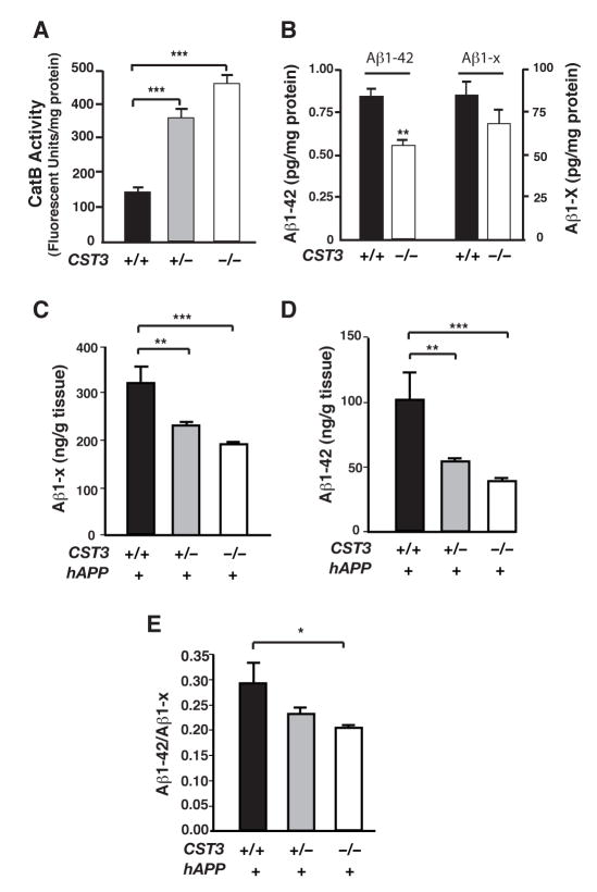
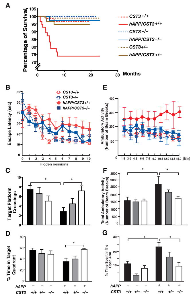
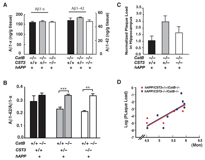
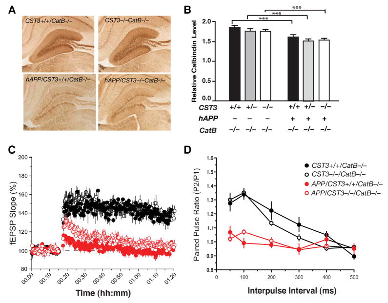
Impaired degradation of amyloid beta (Abeta) peptides could lead to Abeta accumulation, an early trigger of Alzheimer's disease (AD). How Abeta-degrading enzymes are regulated remains largely unknown. Cystatin C (CysC, CST3) is an endogenous inhibitor of cysteine proteases, including cathepsin B (CatB), a recently discovered Abeta-degrading enzyme. A CST3 polymorphism is associated with an increased risk of late-onset sporadic AD. Here, we identified CysC as the key inhibitor of CatB-induced Abeta degradation in vivo. Genetic ablation of CST3 in hAPP-J20 mice significantly lowered soluble Abeta levels, the relative abundance of Abeta1-42, and plaque load. CysC removal also attenuated Abeta-associated cognitive deficits and behavioral abnormalities and restored synaptic plasticity in the hippocampus. Importantly, the beneficial effects of CysC reduction were abolished on a CatB null background, providing direct evidence that CysC regulates soluble Abeta and Abeta-associated neuronal deficits through inhibiting CatB-induced Abeta degradation.


Figures

Figure 1
Figure 1. CysC Reduction Increases CatB Activity and Lowers Soluble Aβ Levels
(A) CatB activities in hippocampal lysates from CST3+/+, CST3+/− or CST3−/− mice (n= 6, ***, P < 0.001, one-way ANOVA with Tukey-Kramer post hoc test). (B) ELISA measurements of Aβ1–42 or Aβ1-x in the supernatants of primary cortical cultures (CST3+/+ or CST3−/−) infected with hAPPV642I adenovirus. All measurements were normalized to protein concentrations in the cell lysates (n= 4, **, P < 0.01, Mann-Whitney U test. Average Aβ levels in CST3+/+ cultures were arbitrarily set as 1). (C–E) ELISA measurements of hippocampal levels of soluble Aβ1-x and Aβ1–42 in 2–4-month-old hAPP-J20 mice. Deleting one or both alleles of CST3 significantly reduced levels of soluble Aβ1-x (C) and Aβ1–42 (D). Ablating CysC also reduced Aβ1–42/Aβ1-x ratios significantly (E). (n = 9–21 mice/genotype, ***, P < 0.001; **, P < 0.01, *, P < 0.05, one-way ANOVA with Tukey-Kramer post hoc test). Bars = means ± SEM (A–E).
Figure 2
Figure 2. CysC Ablation in hAPP-J20 Mice Lowers Total Plaque Deposition
(A and E) Photomicrographs of 3D6 immunostaining (A) or thioflavin S staining (E) in the hippocampus of a 5–8-month-old hAPP/CST3+/+ or hAPP/CST3−/− mouse. (B, D and F) Quantification of plaque load labeled with 3D6 (B and D) or thioflavin S (F) in 5–8-month-old and 8–10-month-old hAPP/CST3+/+ or hAPP/CST3−/− mice (n= 9–15 mice/genotype, **, P < 0.01, *, P < 0.05, unpaired t test). For total plaque load, three hippocampal (B) or cortical (D) sections per mouse were analyzed to determine the percent area covered by 3D6-immunoreactive material. The number of thioflavin S–positive plaques was counted in five hippocampal sections per mouse (F). Bars represent means ± SEM. (C) The logarithm-transformed plaque load was regressed against both age (5–10-month, n= 23–25 mice/genotype) and genotype (as a dummy variable) and was fit to the following model: Log (Plaque Load) = (−7.912 + (0.892) * Age + (1.015) * Genotype (Dummy). The age-adjusted plaque load in hAPP/CST3+/+ mice (Genotype dummy = 1) was significantly higher than that in hAPP/CST3−/− mice (Genotype dummy = 0), since the β2 coefficient (1.015) is significantly larger than 0 (t = 6.217, P < 0.001).
Figure 3
Figure 3. CysC Ablation Does Not Affect the Processing of hAPP
(A) Western blot analyses of cortical lysates demonstrate comparable levels of hAPP metabolic fragments in 5–8-month-old hAPP/CST3+/+ and hAPP/CST3−/− mice. (B–C) Quantification of levels of α-CTFs (B) or α-CTFs (C) relative to those of GAPDH in hAPP/CST3+/+ (n = 9) and hAPP/CST3−/− mice (n = 7). Bars represent means ± SEM. (D) Western blot analyses demonstrate comparable levels of hAPP metabolic fragments in the lysates of CST3+/+ or CST3−/− primary cortical cultures infected with an adenoviral vector expressing wild-type hAPP. GAPDH was used as a loading standard.
Figure 4
Figure 4. CysC Reduction Prevents Functional Deficits in hAPP-J20 Mice
(A) Kaplan-Meier survival curves showing the effect of CysC reduction on survival of hAPP-J20 mice. All genotyped mice in the colony (n = 466) were included in the analysis. By log-rank comparison, only hAPP/CST3+/+ mice differed from all other groups (P < 0.0001). (B–D) Effects of CysC ablation on learning and memory in water maze testing (n= 6–7 male mice/genotype, 7–10 months of age).(B) Hidden platform learning curves differed by genotype (Repeated measure ANOVA: P < 0.0001). In Tukey-Kramer post-hoc comparisons, hAPP/CST3+/+ differed from CST3+/+ and CST3−/− mice (P < 0.001); hAPP/CST3−/− differed from hAPP/CST3+/+ (P < 0.01) but not from CST3+/+ or CST3−/− mice. (C) Number of target platform crossings differed by genotype in the probe trials (Two-way ANOVA, hAPP and CST3 interaction, P < 0.05). In bonferroni post-hoc comparisons, hAPP/CST3+/+ differed from CST3+/+ mice (*, P < 0.05); hAPP/CST3−/− differed from hAPP/CST3+/+ (*, P < 0.05), but not from CST3+/+ or CST3−/− mice. (D) Percentage of time spent in the target quadrant differed by genotype (Two-way ANOVA, hAPP and CST3 interaction, P < 0.05). In bonferroni post-hoc comparisons, hAPP/CST3−/− differed from hAPP/CST3+/+ mice (*, P < 0.05), but not from CST3+/+ or CST3−/− mice. (E–G) Effects of CysC reduction in open field tests and elevated plus maze (n= 5–10 male mice/genotype; 3–4 months of age). (E) The ambulatory activity per min differed by genotype (Repeated-measures ANOVA, P < 0.001). In Tukey-Kramer post-hoc comparisons, hAPP/CST3+/+ differed from CST3+/+ or CST3−/− (P < 0.001), hAPP/CST3+/+ differed from hAPP/CST−/− (P < 0.001). (F) Total ambulatory activity over 15 min differed by genotype (Two-way ANOVA; bonferroni post-hoc comparisons, hAPP/CST3+/+ vs. CST3+/+ *, P < 0.05; hAPP/CST3−/− vs. hAPP/CST3+/+, *, P < 0.05). (G) The percentage of time spent in the open arms of an elevated plus maze differ by genotype (Two-way ANOVA; bonferroni post-hoc comparisons, hAPP/CST3+/+ vs. CST3+/+, *, P < 0.05; hAPP/CST3−/− vs. hAPP/CST3+/+, *, P < 0.05). Error bars represent SEM (B–G).
Figure 5
Figure 5. CysC Ablation Ameliorates Aβ-Associated Neuronal and Synaptic Deficits in Hippocampal Circuits
(A) Photomicrographs of calbindin immunostaining in the hippocampus of a 5–8-month-old hAPP/CST3+/+, hAPP/CST3−/−, CST3+/+, or CST3−/− mouse. (B) Effects of CysC reduction on the relative calbindin levels in the DG (n= 27–46 mice/genotype, age 2–8 months; two-way ANOVA: P < 0.0001, hAPP and CST3 interaction). In post-hoc comparisons, hAPP/CST3+/+ differed from groups without hAPP (***, P < 0.001); hAPP/CST3−/− differed from hAPP/CST3+/+ (***, P < 0.001), but not from all groups without hAPP; hAPP/CST3+/− differed from hAPP/CST3+/+ (*, P < 0.05). CB, cabindin. (C–F) Effects of CysC ablation on the synaptic functions at the perforant path to DG synapses (C–E) or at the Schaffer collateral to CA1 synapse (F) (n=8–12 slices from 2–3 mice/genotype). (C) LTP at the medial perforant path was significantly depressed in hAPP/CST3+/+ mice; deleting CysC in hAPP-J20 mice led to a significant recovery (P < 0.05, CST3+/+ vs. hAPP/CST3+/+; P < 0.05, hAPP/CST3+/+ vs. hAPP/CST3−/−; Tukey’s post-hoc analysis on repeated-measures ANOVA for data collected from minutes 61–70). (D) Paired pulse facilitation at the lateral perforant pathway was markedly reduced in hAPP/CST3+/+ mice; deleting CysC in hAPP-J20 mice led to a significant recovery (P < 0.01, CST3+/+ vs. hAPP/CST3+/+; P < 0.01, hAPP/CST3+/+ vs. hAPP/CST3−/−; One-way ANOVA at 50 and 100 ms interpulse intervals). (E) The synaptic strength at the medial perforant path was not affected by expression of hAPP or deletion of CST3. (F) CysC deletion did not restore the synaptic transmission along the Schaffer collateral-CA1 synapse, which was significantly impaired in hAPP/CST3+/+ mice (P < 0.01, CST3+/+ vs. hAPP/CST3+/+, ANOVA). Error bars represent SEM (B–F).
Figure 6
Figure 6. The Aβ-Lowering Effects of CysC Reduction Depend on CatB
(A) ELISA measurements of levels of soluble Aβ1–42 and Aβ1-x in the hippocampus of 2–3-month-old hAPP/CST3+/+CatB−/−, hAPP/CST3+/−CatB−/−, and hAPP/CST3−/−CatB−/− mice (n= 6–10 mice/genotype). CysC reduction had no effects on the levels of Aβ1-x or Aβ1–42 in hAPP-J20 mice lacking CatB. (B) CatB ablation increased the ratios of Aβ1–42/Aβ1-x in hAPP-J20 mice with one or both alleles of CST3 deleted (n= 6–21 mice/genotype, age 2–4 months; two-way ANOVA: ***, P < 0.001, **, P < 0.01, bonferroni post-hoc test of the CatB effects). (C) Normalized plaque load in hippocampus of 4–6-month-old hAPP/CST3+/+CatB−/−, hAPP/CST3+/−CatB−/−, and hAPP/CST3−/−CatB−/− mice (n= 7–14 mice/genotype; one-way ANOVA, P > 0.1). (D) The logarithm-transformed plaque load was regressed against both age and genotype (as a dummy variable) and was fit to the following model: Log (Plaque Load) = (−14.547) + (2.052) * Age + (−0.111) * Genotype (Dummy). The age-adjusted plaque load in hAPP/CST3+/+CatB−/− mice (Genotype dummy = 1) did not differ from that in hAPP/CST3−/−CatB−/− mice (Genotype dummy = 0), since the β2 coefficient (−0.111) is not significantly different from 0 (t = −0.258, P = 0.8).
Figure 7
Figure 7. The Neuroprotective Effects of CysC Reduction Depend on CatB
(A) Photomicrographs of calbindin immunostaining in the hippocampus of 5-month-old hAPP/CST3+/+CatB−/−, hAPP/CST3−/−CatB−/−, CST3+/+CatB−/−, and CST3−/−CatB−/−, mice. (B) CysC reduction failed to prevent the depletion of DG calbindin levels in hAPP-J20 mice lacking CatB (n= 9–22 mice/genotype, age 2–6 months old; two-way ANOVA: ***, P < 0.001, bonferroni post-hoc test of the hAPP effects; CST3 effects, not significant). (C–D) Effects of CysC ablation on the synaptic functions in hippocampus of hAPP-J20 mice on CatB null background (n=8–12 slices from 2 mice/genotype). (C) LTP at the medial perforant path was significantly depressed in hAPP/CatB−/− mice regardless of CST3 genotypes (P < 0.01, CST3+/+CatB−/− vs. hAPP/CST3+/+CatB−/− and CST3−/−CatB−/− vs. hAPP/CST3−/−CatB−/−). CysC removal did not alter LTP (P > 0.1, hAPP/CST3+/+CatB−/− vs. hAPP/CST3−/−CatB−/−; Tukey’s post-hoc analysis on repeated-measures ANOVA for data collected from minutes 61–70). (D) The paired pulse facilitations at the lateral perforant pathway were markedly reduced in hAPP/CatB−/− mice regardless of CST3 genotypes (P < 0.01, CST3+/+CatB−/− vs. hAPP/CST3+/+CatB−/− ; CST3−/−CatB−/− vs. hAPP/CST3−/−CatB−/−; One-way ANOVA at 50–100 ms interpulse intervals). Bars represent means ± SEM (B–D).

References

    1. Abrahamson M, Barrett AJ, Salvesen G, Grubb A. Isolation of six cysteine proteinase inhibitors from human urine. Their physicochemical and enzyme kinetic properties and concentrations in biological fluids. J Biol Chem. 1986;261:11282–11289. - PubMed
    1. Benussi L, Ghidoni R, Steinhoff T, Alberici A, Villa A, Mazzoli F, Nicosia F, Barbiero L, Broglio L, Feudatari E, et al. Alzheimer disease-associated cystatin C variant undergoes impaired secretion. Neurobiol Dis. 2003;13:15–21. - PubMed
    1. Bertram L, McQueen MB, Mullin K, Blacker D, Tanzi RE. Systematic meta-analyses of Alzheimer disease genetic association studies: the AlzGene database. Nat Genet. 2007;39:17–23. - PubMed
    1. Beyer K, Lao JI, Gomez M, Riutort N, Latorre P, Mate JL, Ariza A. Alzheimer’s disease and the cystatin C gene polymorphism: An association study. Neurosci Lett. 2001;315:17–20. - PubMed
    1. Bogyo M, Verhelst S, Bellingard-Dubouchaud V, Toba S, Greenbaum D. Selective targeting of lysosomal cysteine proteases with radiolabeled electrophilic substrate analogs. Chem Biol. 2000;7:27–38. - PubMed

Publication types