. 2008 Oct 23;60(2):321-7.
doi: 10.1016/j.neuron.2008.08.026.

Reverberation of recent visual experience in spontaneous cortical waves

Affiliations

Reverberation of recent visual experience in spontaneous cortical waves

Feng Han et al. Neuron. .

Abstract

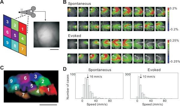
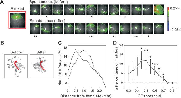
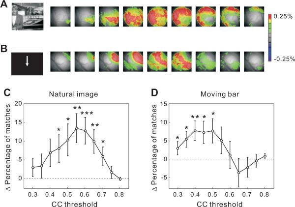
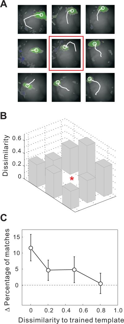
Spontaneous waves of activity propagating across large cortical areas may play important roles in sensory processing and circuit refinement. However, whether these waves are in turn shaped by sensory experience remains unclear. Here we report that visually evoked cortical activity reverberates in subsequent spontaneous waves. Voltage-sensitive dye imaging in rat visual cortex shows that following repetitive presentation of a given visual stimulus, spatiotemporal activity patterns resembling the evoked response appear more frequently in the spontaneous waves. This effect is specific to the response pattern evoked by the repeated stimulus, and it persists for several minutes without further visual stimulation. Such wave-mediated reverberation could contribute to short-term memory and help to consolidate the transient effects of recent sensory experience into long-lasting cortical modifications.


Figures

Figure 1
Figure 1
Spontaneous and evoked waves in rat visual cortex (A) Schematic illustration of visual stimulation and cranial window for VSD imaging. Positions for visual stimulation are marked with numbers and colors (left). For the image of the visual cortex (right), the left-right axis corresponds to anterior-posterior axis of the cortex; the upper-lower axis corresponds to lateral-medial. This imaging area contains mostly V1 and a small portion of V2 (lower left region). Scale bar, 1 mm. (B) Examples of spontaneous and visually evoked waves, shown in 20 ms intervals. VSD signal was color coded (warm color indicates depolarization), with scale bar shown on the right. Top, two examples of spontaneous waves. Bottom, waves evoked by stimuli at positions 1 & 9 (average of 2 trials; 7 frames following stimulus onset but before response onset were omitted). White circle, initiation site measured as center of mass of VSD signal in the first frame. White line, wave trajectory by connecting the centers of mass of consecutive frames. Across experiments the mean amplitude was not significantly different between spontaneous and evoked waves (p > 0.9, Mann-Whitney U test; evoked: ΔF/F = 0.231 ± 0.046%, SD; spontaneous: ΔF/F = 0.234 ± 0.051%). (C) Initiation sites of waves evoked by the nine stimuli (retinotopy), with colors and numbers corresponding to (A). Scale bar, 1mm. (D) Distributions of the instantaneous wave speed, measured as the distance in the center of mass of ΔF/F between consecutive frames divided by the inter-frame interval (10 ms). For each wave, up to 18 instantaneous speeds were measured. Shown are data from 9 rats; in each rat, the stimuli were flashed at multiple positions to measure the evoked waves.
Figure 2
Figure 2
Effect of repeated visual stimulation at a given position on spontaneous waves (A) Spontaneous waves immediately before and after training (10.24 s/session). Each image shows the initial frame of a wave. Initiation site and propagation path are indicated by a white circle and a line. Left, Evoked wave in response to the training stimulus. Spontaneous waves well matched to the template are indicated; ▲, CC>0.6; ▲▲, CC>0.7. (B) Superposition of the initiation sites and propagation paths for all spontaneous waves before and after training (gray) and for the training-evoked wave (red). Dotted circle indicates a 0.6 mm radius from the initiation site of the training-evoked wave. (C) Distribution of the distance between the initiation sites of the spontaneous waves and the evoked template before (dashed) and after (solid) training (30 training experiments, 9 rats). (D) Difference in the percentage of matched waves before and after training plotted as a function of the CC threshold (*, p < 0.05; **, p < 0.02; ***, p < 0.01, same data as in C). Error bar, ± SEM.
Figure 3
Figure 3
Effect of repeated presentation of natural images and moving bars on spontaneous waves (A) Example of a wave evoked by a natural image, shown in 20 ms intervals. Left, The natural image used for stimulation. VSD signal was color coded as in Figure 1 (average of 3 trials; 7 frames following stimulus onset but before response onset were omitted). (B) Example of a wave evoked by a moving bar, shown in 20 ms intervals (average of 3 trials; 7 frames following stimulus onset but before response onset were omitted). (C) Difference in the percentage of matched spontaneous waves before and after training with natural images plotted as a function of CC threshold (*, p < 0.05; **, p < 0.005; ***, p < 0.001, 38 training experiments, 5 rats). (D) Same as (A), for moving bars as training stimuli (49 training experiments, 19 rats).
Figure 4
Figure 4
Specificity of the effect to the training pattern after 50 repeats of flashed squares at 0.6 Hz (A) Initiation sites and trajectories of waves evoked by the nine stimuli in an example experiment. Red box indicates response to the training stimulus. (B) Dissimilarity (1-CC) between the wave evoked by each non-training stimulus and that evoked by the training stimulus for the experiment shown in (A). Red star, dissimilarity for the training stimulus (=0). (C) Change in the percentage of matches between spontaneous waves and all evoked templates plotted against the dissimilarity between the untrained and trained templates. Data are from the same 30 experiments as in Figure 2. Error bar, ± SEM.
Figure 5
Figure 5
Induction and decay time course of the effect induced by flashed squares (A) Change in the percentage of matches immediately after training vs. number of training stimuli (10 repeats: 24 training experiments, 8 rats; 25 repeats: 28 experiments, 8 rats; 50 repeats: 30 experiments, 9 rats; 125 repeats: 44 experiments, 16 rats). (B) Persistence of ΔCC induced by 50 (gray) and 125 (black) training stimuli (30 and 44 training experiments for gray and black lines, respectively).

References

    1. Abeles M, Gerstein GL. Detecting spatiotemporal firing patterns among simultaneously recorded single neurons. J Neurophysiol. 1988;60:909–924. - PubMed
    1. Amzica F, Steriade M. Short- and long-range neuronal synchronization of the slow (< 1 Hz) cortical oscillation. J Neurophysiol. 1995;73:20–38. - PubMed
    1. Arieli A, Sterkin A, Grinvald A, Aertsen A. Dynamics of ongoing activity: explanation of the large variability in evoked cortical responses. Science. 1996;273:1868–1871. - PubMed
    1. Averbach E, Sperling G. Short term storage of information in vision. In: Cherry C, editor. Information Theory. Butterworth; London: 1961. pp. 196–211.
    1. Benucci A, Frazor RA, Carandini M. Standing waves and traveling waves distinguish two circuits in visual cortex. Neuron. 2007;55:103–117. - PMC - PubMed

Publication types

Substances

LinkOut - more resources