. 2008 Oct 23;60(2):378-89.
doi: 10.1016/j.neuron.2008.09.023.

Integrating memories in the human brain: hippocampal-midbrain encoding of overlapping events

Affiliations

Integrating memories in the human brain: hippocampal-midbrain encoding of overlapping events

Daphna Shohamy et al. Neuron. .

Abstract

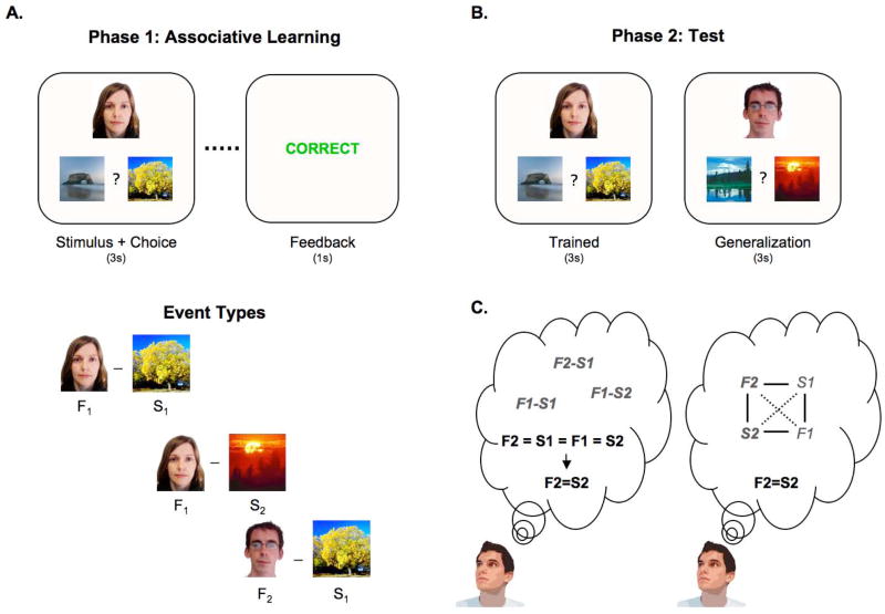
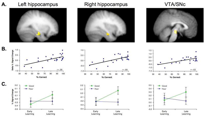
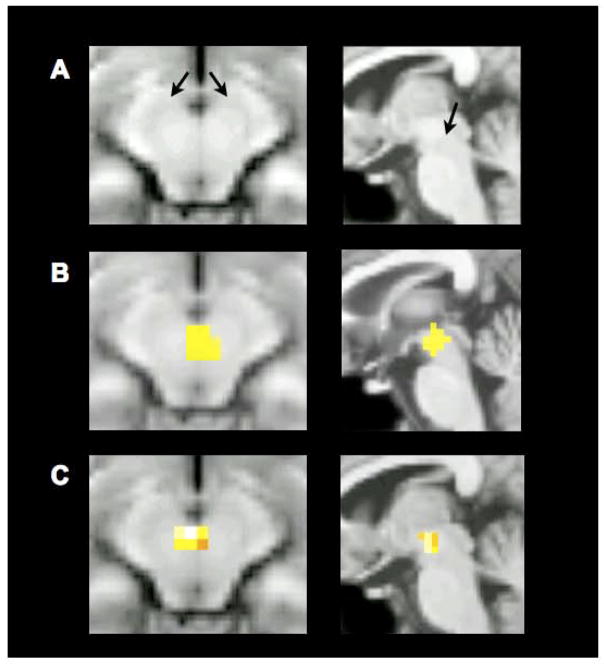
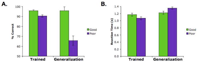
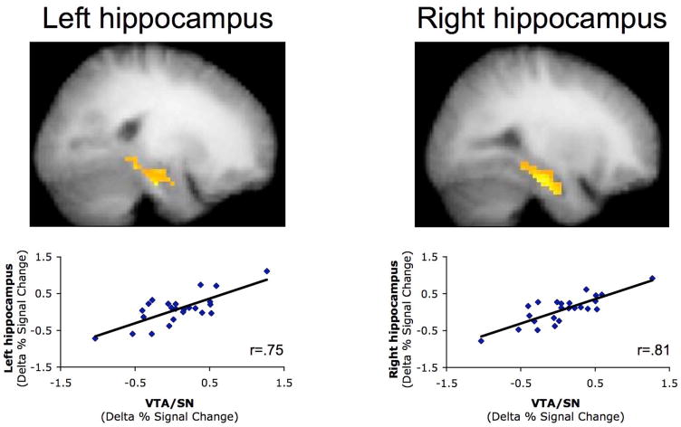
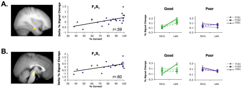
Decisions are often guided by generalizing from past experiences. Fundamental questions remain regarding the cognitive and neural mechanisms by which generalization takes place. Prior data suggest that generalization may stem from inference-based processes at the time of generalization. By contrast, generalization may emerge from mnemonic processes occurring while premise events are encoded. Here, participants engaged in a two-phase learning and generalization task, wherein they learned a series of overlapping associations and subsequently generalized what they learned to novel stimulus combinations. Functional MRI revealed that successful generalization was associated with coupled changes in learning-phase activity in the hippocampus and midbrain (ventral tegmental area/substantia nigra). These findings provide evidence for generalization based on integrative encoding, whereby overlapping past events are integrated into a linked mnemonic representation. Hippocampal-midbrain interactions support the dynamic integration of experiences, providing a powerful mechanism for building a rich associative history that extends beyond individual events.


Figures

Figure 1
Figure 1
Representative events and structure of the task. (A) Participants learned a series of individual face-scene associations based on feedback (36 individual associations in total). On each trial, the face-scene pair was presented for 3 s, after which performance-dependent feedback was provided for 1 s. There were three learning event types—the individual associations shared overlapping features, with two faces always associated with a common scene, and one of those faces also associated with a second scene. A scene that was the incorrect choice for one face was the correct choice for another face, so that simple stimulus-response learning strategies could not support learning. (B) After learning, participants underwent a test phase, where they received no feedback and where they were asked to respond to untrained face-scene associations. These generalization trials were presented together with trials that tested knowledge for previously trained associations. (C) The generalization trials can be correctly responded to by way of two different mechanisms: during test, retrieval of the previously trained individual associations may allow participants to draw inferences across them (left); alternatively, the untrained association may have been formed during learning due to retrieval and integrative encoding that is triggered by the overlapping features across individual trained associations.
Figure 2
Figure 2
Hippocampal and midbrain activation during learning predicts correct responding on the generalization trials at test. (A) Map-wise regression analyses revealed that the change in activation from early to late learning in left hippocampus (−28,−9,−17; 27 voxels), right hippocampus (31,−5,−20; 22 voxels), and a bilateral midbrain complex (3,−18,−12; 50 voxels) correlated with % correct generalization performance at test (P<0.001, extent threshold 5 voxels; P<0.05, small volume corrected for the hippocampus and midbrain). (B) BOLD % signal change data extracted from these hippocampal and midbrain regions (inclusive of all above-threshold voxels within a 6-mm sphere surrounding the peak voxel) confirmed the strong correlation between learning-phase activation increases and generalization performance. (C) When participants were median split based on generalization performance at test, an increase in hippocampal and midbrain activation from early to late learning was observed in participants who generalized well (‘good’ group), but not in participants who generalized poorly (‘poor’ group). Error bars +/− S.E.M.
Figure 3
Figure 3
Localization of midbrain activations, displayed on a canonical T1-weighted image (axial slice, left; sagittal slice, right). The midbrain complex consists of the substantia nigra (SN) and the ventral tegmental area (VTA). (A) SN extends lateral and posterior around the oval red nuclei, as indicated by the black arrows. VTA is medial to SN, and borders the interpeduncular cistern. (B) Higher magnification of the generalization-related midbrain region-of-interest described in the main findings (data smoothed with an 8-mm filter; P<0.001, extent threshold 5 voxels; P<0.05, small volume corrected for the hippocampus and midbrain). (C) Visualization of generalization-related midbrain activations revealed when using a smaller (4-mm) smoothing filter during functional data preprocessing (P<0.001, extent threshold 5 voxels; P<0.05, small volume corrected for the hippocampus and midbrain). Full reporting of the data smoothed with a 4-mm filter appear in the Supplemental Results.
Figure 4
Figure 4
Behavioral performance at test on trained and generalization trials for the ‘good’ and ‘poor’ generalization participants. (A) An interaction between group and test trial type revealed a significantly greater difference between the two groups in performance on the generalization trials relative to the trained trials. Importantly, the ‘good’ group showed no difference in accuracy between trained and generalization trials, whereas the ‘poor’ group showed superior performance on trained than on generalization trials. (B) This pattern was also evident in the speed of responding. The ‘poor’ group, relative to the ‘good’ group, showed a marked difference in response latencies to trained vs. generalization trials, consistent with the hypothesis that in the ‘good’ group the associations necessary to rapidly respond to generalization trials were constructed during learning. Error bars +/− S.E.M.
Figure 5
Figure 5
Learning-phase activation changes in bilateral hippocampus demonstrated a significant correlation with such changes in the midbrain. This analysis regressed the difference in % signal change from early to late learning in a seed region in the midbrain with voxels in the medial temporal lobe (P<0.001, extent threshold 5 voxels; small volume corrected, P<0.05). Extracting the change in integrated % signal change for all activated voxels in left and right hippocampus (98 and 161 voxels, respectively) identified in this regression confirmed the tight relationship with the change in integrated % signal change in the midbrain. Importantly, this relationship between hippocampal and midbrain activation remained significant even when excluding the two participants demonstrating the strongest and weakest change in midbrain learning-phase activity (left hippocampus–midbrain r=0.59; P<0.005; right hippocampus–midbrain, r=0.75, P<0.001).
Figure 6
Figure 6
Hippocampal and midbrain activation to different event types during learning. The relationship between (A) hippocampal and (B) midbrain activation during learning and generalization performance at test was strongest for the F2–S1 learning trials. Regression analyses (left) revealed that subsequent generalization correlated with learning-phase activation increases to F2–S1 trials in both bilateral hippocampus (data shown for right hippocampus) and midbrain (Ps<.05, corrected); no other correlations survived correction for multiple comparisons. Similarly, increased activation during learning of F2–S1 events showed the strongest difference across ‘good’ and ‘poor’ generalization subgroups. The ‘good’ (Ps<0.05), but not the ‘poor’ (Ps>0.40), generalization group demonstrated a significant increase in bilateral hippocampal and midbrain activation from early to late learning of F2–S1 trials. In the midbrain, this increase was selective to the F2–S1 trials, whereas in the hippocampus, a qualitatively similar effect was observed for the F1–S1 and F1–S2 trials (see Supplemental Results). Error bars +/− S.E.M.

Comment in

References

    1. Adcock RA, Thangavel A, Whitfield-Gabrieli S, Knutson B, Gabrieli JD. Reward-motivated learning: mesolimbic activation precedes memory formation. Neuron. 2006;50:507–517. - PubMed
    1. Aron AR, Shohamy D, Clark J, Myers C, Gluck MA, Poldrack RA. Human midbrain sensitivity to cognitive feedback and uncertainty during classification learning. J Neurophysiol. 2004;92:1144–1152. - PubMed
    1. Bayer HM, Glimcher PW. Midbrain dopamine neurons encode a quantitative reward prediction error signal. Neuron. 2005;47:129–141. - PMC - PubMed
    1. Bunzeck N, Duzel E. Absolute coding of stimulus novelty in the human substantia nigra/VTA. Neuron. 2006;51:369–379. - PubMed
    1. Cohen NJ, Eichenbaum H. Memory, amnesia, and the hippocampal system. Cambridge, MA: MIT Press; 1993.

Publication types

MeSH terms