

Review
doi: 10.1016/j.sbi.2008.09.010.
Epub 2008 Nov 17.
Structural insights into G-protein-coupled receptor activation
Affiliations
- PMID: 18957321
- PMCID: PMC4019673
- DOI: 10.1016/j.sbi.2008.09.010
Item in Clipboard
Review
Structural insights into G-protein-coupled receptor activation
Curr Opin Struct Biol.
2008 Dec.
Abstract
G-protein-coupled receptors (GPCRs) are the largest family of eukaryotic plasma membrane receptors, and are responsible for the majority of cellular responses to external signals. GPCRs share a common architecture comprising seven transmembrane (TM) helices. Binding of an activating ligand enables the receptor to catalyze the exchange of GTP for GDP in a heterotrimeric G protein. GPCRs are in a conformational equilibrium between inactive and activating states. Crystallographic and spectroscopic studies of the visual pigment rhodopsin and two beta-adrenergic receptors have defined some of the conformational changes associated with activation.
Figures
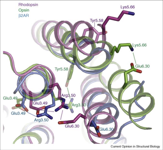
An overlay of dark rhodopsin (purple; PDB 1gzm), low pH opsin (green; PDB 3cap), and carazolol-bound β2AR–T4L (blue; PDB 2rh1) in the vicinity of the ionic lock. In dark rhodopsin, Arg1353.50 in the conserved D/ERY sequence near the cytoplasmic end of TM3 forms a salt bridge with Glu2476.30 at the cytoplasmic end of TM6. Arg3.50 is further stabilized by a salt bridge to the preceding conserved acid at position 3.49 in both rhodopsin and β2AR. In opsin, Arg1353.50 interacts with Tyr2235.58 in TM5, and Glu2476.30 forms a salt bridge with Lys2315.66. The homologous ionic lock residues Arg1313.50 and Glu2686.30 from the β2AR are also shown. The amino acids are numbered using the Ballesteros–Weinstein system. Within each helix is a single most conserved residue among the class A GPCRs. This residue is designated x.50, where x is the number of the transmembrane helix. All other residues on that helix are numbered relative to this conserved position.

Comparison of rhodopsin (purple; PDB 1gzm) and carazolol-bound β2AR–T4L (blue; PDB 2rh1). Extracellular loop 2 (ECL2) of rhodopsin forms a lid over the retinal-binding pocket, whereas the position of ECL2 of the β2AR allows relatively free access to the carazolol-binding pocket.

Superimposed structures of rhodopsin (purple; PDB 1gzm) and carazolol-bound β2AR–T4L (blue; PDB 2rh1) highlighting water molecules that form a hydrogen-bond network with conserved amino acids. Atoms of carazolol and retinal are shown as spheres. The amino acids are numbered using the Ballesteros–Weinstein system (see Figure 1).
References
-
- Kobilka BK, Deupi X. Conformational complexity of G-protein-coupled receptors. Trends Pharmacol Sci. 2007;28:397–406. - PubMed
-
- Krebs A, Villa C, Edwards PC, Schertler GF. Characterisation of an improved two-dimensional p22121 crystal from bovine rhodopsin. J Mol Biol. 1998;282:991–1003. - PubMed
-
- Schertler GF, Villa C, Henderson R. Projection structure of rhodopsin. Nature. 1993;362:770–772. - PubMed
-
- Palczewski K, Kumasaka T, Hori T, Behnke CA, Motoshima H, Fox BA, Le Trong I, Teller DC, Okada T, Stenkamp RE, et al. Crystal structure of rhodopsin: a G protein-coupled receptor [see comments]. Science. 2000;289:739–745. - PubMed
-
- Okada T, Le Trong I, Fox BA, Behnke CA, Stenkamp RE, Palczewski K. X-ray diffraction analysis of three-dimensional crystals of bovine rhodopsin obtained from mixed micelles. J Struct Biol. 2000;130:73–80. - PubMed
Publication types
MeSH terms
Substances
Grants and funding
LinkOut - more resources
Full Text Sources
