. 2009 Jan 27;364(1514):209-15.
doi: 10.1098/rstb.2008.0126.

Proton-coupled protein transport through the anthrax toxin channel

Affiliations

Proton-coupled protein transport through the anthrax toxin channel

Alan Finkelstein. Philos Trans R Soc Lond B Biol Sci. .

Abstract

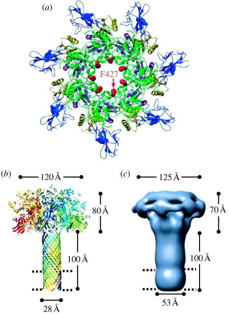
Anthrax toxin consists of three proteins (approx. 90kDa each): lethal factor (LF); oedema factor (OF); and protective antigen (PA). The former two are enzymes that act when they reach the cytosol of a targeted cell. To enter the cytosol, however, which they do after being endocytosed into an acidic vesicle compartment, they require the third component, PA. PA (or rather its proteolytically generated fragment PA63) forms at low pH a heptameric beta-barrel channel, (PA63)7, through which LF and OF are transported--a phenomenon we have demonstrated in planar phospholipid bilayers. It might appear that (PA63)7 simply forms a large hole through which LF and OF diffuse. However, LF and OF are folded proteins, much too large to fit through the approximately 15A diameter (PA63)7 beta-barrel. This paper discusses how the (PA63)7 channel both participates in the unfolding of LF and OF and functions in their translocation as a proton-protein symporter.


Figures

Figure 1
Figure 1
Structure of the (PA63)7 pore (channel) and pre-pore. (a) Ribbons rendering of the pre-pore viewed axially. The phenylalanines at residue 427 are seen to face the lumen of the pre-pore. After conversion of the pre-pore to the pore, they continue to face the channel lumen, but, as evidenced from EPR spectra of spin label introduced uniformly at this site (Krantz et al. 2005), they have moved closer together; that is, the lumen diameter is smaller (adapted from Krantz et al. 2005). (b) Model of the mushroom-like (PA63)7 channel based on the crystal structure of monomeric PA, the crystal structure of the heptameric α-haemolysin channel and cysteine mutagenesis experiments that established the length of the β-barrel stem. The ring of phenylalanines at residue 427 (the ϕ-clamp) lies near the junction of the cap and the stem. The parallel dashed lines here and in (c) indicate the thickness of the hydrophobic interior of the phospholipid bilayer (adapted from Nguyen 2004, published with permission from Adenine Press, Inc. http://www.jbsdonline.com). (c) The structure of the (PA63)7 channel as obtained from negative-stain electron microscopy at a resolution of approximately 25 Å. The agreement of this structure with the model in (b) is gratifying (adapted from Katayama et al. 2008, adapted with permission from Macmillan Publishers Ltd; see http://www.nature.com/nsmb).
Figure 2
Figure 2
The interaction of LFN with the (PA63)7 channel. After the (PA63)7-induced conductance had reached a more-or-less steady state, LFN was added (at the arrow) to the cis side to a concentration of 6 nM, resulting in a rapid fall in conductance. LFN (along with (PA63)7) was then perfused out of the cis compartment (during the approx. 4 min break in the record); the conductance increased only slightly over this time. When the voltage was stepped from +20 to −20 mV, there was a very rapid increase in conductance followed by a slower increase. When the voltage was stepped back to +20 mV, the conductance rapidly fell to a value somewhat larger than before. By contrast, when the voltage was stepped from +20 to +45 mV, there was an S-shaped rise of conductance to a value comparable to that before the addition of LFN, and it remained at that value when the voltage was stepped back from +45 to +20 mV.
Figure 3
Figure 3
Cartoon of the interaction of LFN with the (PA63)7 channel and the effect of voltage and a pH gradient on this interaction. (The distribution of positively and negatively charged groups in the 263 residues of LFN is shown at the top.) The (PA63)7 channel is diagrammed as a long narrow stem, the bottom third of which crosses the bilayer (represented by the parallel dashed lines), opening up into a wide vestibule. The constriction near the junction of the vestibule with the stem is the ϕ-clamp formed by the seven F427s. At the top of the vestibule is a binding site for LFN; LFN is schematized as a folded structure with the N-terminal 26 residues disordered (Pannifer et al. 2001) and therefore able to enter the channel and block it at +20 mV once the N-terminal end has traversed the ϕ-clamp. The N-terminal end is driven out of the channel at −20 mV and thereby unblocks it, but remains attached to its binding site and only slowly dissociates from this site into the cis solution. (The degree of penetration of LFN into the channel at +20 mV has not yet been determined, and conceivably the N-terminal end may even traverse the entire channel and emerge into the trans solution.) At larger positive voltages (+45 mV), LFN unfolds with the aid of the ϕ-clamp (see text), and its extended structure is driven through the stem until ultimately the entire LFN protein has been translocated across the membrane. Before entering the cation-selective stem, the carboxyl groups on glutamates and aspartates pick up protons from the cis side and are thereby neutralized; thus, the LFN segment within the stem always bears a positive charge (or at worst is neutral) and is consequently driven by the electric field from the cis to the trans side. (The protonation of the aspartates and glutamates is indicated as occurring before they cross the ϕ-clamp, but it is possible that it occurs at some point after this in the stem.) As they exit the channel, these carboxyl groups release their protons into the trans solution, completing the co-transport of protons with the protein. Alternatively, instead of the translocation being driven by a step of voltage from +20 to +45 mV, it can be driven by raising the trans pH (see text). Blue circles, Arg+/His+/Lys+; green circles, H+; yellow circles, Glu0/Asp0; red circles, Glu/Asp. This figure is an elaboration of one in Zhang et al. (2004b).
Figure 4
Figure 4
LFN translocation being driven by a pH gradient. Prior to the start of the record, the (PA63)7-induced conductance was reduced over 30-fold by the addition (at symmetric pH 5.5 and +20 mV) of LFN to the cis solution, after which LFN was then perfused out of that solution. We see that raising the trans pH to 5.85 induced a slow rate of rise in conductance, reflecting the translocation of LFN, and raising it further to 6.2 caused a much greater increase in the rate of LFN translocation (adapted from Krantz et al. 2006).

References

    1. Blaustein R.O., Finkelstein A. Voltage-dependent block of anthrax toxin channels in planar phospholipid bilayer membranes by symmetric tetraalkylammonium ions: effects on macroscopic conductance. J. Gen. Physiol. 1990a;96:905–919. doi:10.1085/jgp.96.5.905 - DOI - PMC - PubMed
    1. Blaustein R.O., Finkelstein A. Diffusion limitation in the block by symmetric tetraalkylammonium ions of anthrax toxin channels in planar phospholipid bilayer membranes. J. Gen. Physiol. 1990b;96:943–957. doi:10.1085/jgp.96.5.943 - DOI - PMC - PubMed
    1. Cabiaux V., Mindell J., Collier R.J. Membrane translocation and channel-forming activities of diphtheria toxin are blocked by replacing isoleucine 364 with lysine. Infect. Immun. 1993;61:2200–2202. - PMC - PubMed
    1. Falnes P.O., Sandvig K. Penetration of protein toxins into cells. Curr. Opin. Cell Biol. 2000;12:407–413. doi:10.1016/S0955-0674(00)00109-5 - DOI - PubMed
    1. Falnes P.O., Madshas I.H., Sandvig K., Olsnes S. Replacement of negative by positive charges in the presumed membrane-inserted part of diphtheria toxin B fragment. Effect on membrane translocation and on formation of cation channels. J. Biol. Chem. 1992;267:12 284–12 290. - PubMed

Publication types