. 2009 Jan 9;284(2):1064-74.
doi: 10.1074/jbc.M807273200. Epub 2008 Oct 28.

Analysis of histones in Xenopus laevis. I. A distinct index of enriched variants and modifications exists in each cell type and is remodeled during developmental transitions

Affiliations

Analysis of histones in Xenopus laevis. I. A distinct index of enriched variants and modifications exists in each cell type and is remodeled during developmental transitions

David Shechter et al. J Biol Chem. .

Abstract

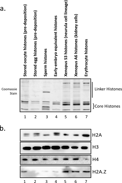
Histone proteins contain epigenetic information that is encoded both in the relative abundance of core histones and variants and particularly in the post-translational modification of these proteins. We determined the presence of such variants and covalent modifications in seven tissue types of the anuran Xenopus laevis, including oocyte, egg, sperm, early embryo equivalent (pronuclei incubated in egg extract), S3 neurula cells, A6 kidney cells, and erythrocytes. We first developed a new robust method for isolating the stored, predeposition histones from oocytes and eggs via chromatography on heparin-Sepharose, whereas we isolated chromatinized histones via conventional acid extraction. We identified two previously unknown H1 isoforms (H1fx and H1B.Sp) present on sperm chromatin. We immunoblotted this global collection of histones with many specific post-translational modification antibodies, including antibodies against methylated histone H3 on Lys(4), Lys(9), Lys(27), Lys(79), Arg(2), Arg(17), and Arg(26); methylated histone H4 on Lys(20); methylated H2A and H4 on Arg(3); acetylated H4 on Lys(5), Lys(8), Lys(12), and Lys(16) and H3 on Lys(9) and Lys(14); and phosphorylated H3 on Ser(10) and H2A/H4 on Ser(1). Furthermore, we subjected a subset of these histones to two-dimensional gel analysis and subsequent immunoblotting and mass spectrometry to determine the global remodeling of histone modifications that occurs as development proceeds. Overall, our observations suggest that each metazoan cell type may have a unique histone modification signature correlated with its differentiation status.


Figures

FIGURE 1.
FIGURE 1.
Isolation of histones from Xenopus tissue and remodeling in egg extract. a, schematic diagram of different cell types used in this study. We pooled oocytes from stages II–VI and separately pooled laid eggs. We subsequently prepared soluble extract and then applied the extract to heparin-Sepharose to isolate purified histones. b, Coomassie-stained gel showing the applied and eluted fractions of proteins from egg extract applied to heparin-Sepharose. The location of the core histones is indicated. Note that Xenopus egg and early embryo histones H2A, H2B, and H3 tend to migrate together in a single band on a high percentage SDS-polyacrylamide gel. c, Western blot of the fractions from the heparin-Sepharose column, showing the retention and specific elution of histones H2A, H2B, H3, and H4. d, Coomassie-stained gel of acid-extracted histones from sperm chromatin (first lane) and sperm chromatin assembled in egg extract (so-called pronuclei chromatin) (second lane). The location of the core histones is noted. Also indicated are the identification of proteins in gel slices from each lane, as determined by mass spectrometry, including the sperm-specific histone H1fx as well as the sperm-specific basic protein 4. Proteins that are acid-soluble and assembled on chromatin during pronucleus production in egg extract were identified by mass spectrometry. Those protein names are listed below the gel. e, Coomassie-stained gel of acid-extracted histones from erythrocyte chromatin (first lane) and erythrocyte chromatin assembled in egg extract (second lane). The location of the core histones is noted, as is the putative location of the somatic H1 types, although these were not positively identified by mass spectrometry. f, listing of mass spectrometry-identified proteins from the indicated gel slice in d.
FIGURE 2.
FIGURE 2.
Isolation and relative enrichment of histone proteins and histone variants in embryonic and somatic cells. a, Coomassie-stained gel of histones isolated from the chaperone-complexed stored population in oocytes and eggs and the acid-extractable chromatin-associated sperm histones, early embryo equivalent (pronuclei) histones, S3 tissue culture histones, A6 tissue culture histones, and erythrocyte histones. The position of the core histones is noted as well as the putative location of the variety of histone H1 proteins in each cell type. b, Western blots of the histones isolated in 2a using antibodies against histone H3, H4, H2A, and H2A.Z.
FIGURE 3.
FIGURE 3.
Relative enrichment of histone post-translation modifications in embryonic and somatic cells. a, a cropped view of the Coomassie-stained gel from the isolated histones in a presented to show the core histones for reference and alignment with the blots in this figure. b, Western blots of the isolated histones with antibodies specific for a range of specific lysine methylation states, as indicated beside each blot. c, Western blots of the isolated histones with antibodies specific for a range of specific lysine acetylation states, as indicated beside each blot. d, Western blots of the isolated histones with antibodies specific for a range of specific arginine methylation states, as indicated beside each blot. e, Western blot of the isolated histones with an antibody against phosphorylated Ser1 of H2A and H4.
FIGURE 4.
FIGURE 4.
Two-dimensional TAU/SDS gel analysis demonstrating remodeling of histones during development. a, Coomassie-stained two-dimensional gel run with isolated Xenopus egg histones. The numbered stained gel bands were cut out and subject to mass spectrometry identification. The identified proteins are listed below the gel. b, Coomassie-stained two-dimensional gel run with isolated Xenopus sperm histones. c, Coomassie-stained two-dimensional gel run with isolated Xenopus pronuclei histones (early embryo equivalent histones). The numbered stained gel bands were cut out and subjected to mass spectrometry for identification. The identified proteins are listed below the gel. d, Coomassie-stained two-dimensional gel run with isolated Xenopus erythrocyte histones. The numbered stained gel bands were cut out and subjected to mass spectrometry for identification. The identified proteins are listed below the gel.
FIGURE 5.
FIGURE 5.
Immunoblots of two-dimensional TAU/SDS gels from early embryo equivalent (pronuclei) histones. a, two-dimensional gel run with isolated Xenopus pronuclei histones. The gel was immunoblotted with an antibody specific for histone H2A; this antibody will identify most H2A isoforms, since it recognizes the acidic patch in the histone fold region. b, two-dimensional gel run with isolated Xenopus pronuclei histones. The gel was immunoblotted with an antibody specific for histone H3. c, two-dimensional gel run with isolated Xenopus pronuclei histones. The gel was immunoblotted with an antibody specific for histone H4. d, two-dimensional gel run with isolated Xenopus pronuclei histones. The gel was immunoblotted with an antibody specific for phosphorylated Ser1 of histones H2A and H4. e, purified histones from egg, sperm, early embryo equivalent pronuclei, and erythrocytes were run on a 17% SDS-polyacrylamide gel and blotted for H2A and the embryonic H2A.X. The modified, mass-shifted, H2A isoform is indicated on the blot.

References

    1. Strahl, B. D., and Allis, C. D. (2000) Nature 403 41-45 - PubMed
    1. Malik, H. S., and Henikoff, S. (2003) Nat. Struct. Biol. 10 882-891 - PubMed
    1. Bernstein, E., and Hake, S. B. (2006) Biochem. Cell Biol. 84 505-517 - PubMed
    1. Ruthenburg, A., Li, H., Patel, D., and Allis, C. (2007) Nat. Rev. Mol. Cell Biol. 8 983-994 - PMC - PubMed
    1. Taverna, S., Li, H., Ruthenburg, A., Allis, C., and Patel, D. (2007) Nat. Struct. Mol. Biol. 14 1025-1040 - PMC - PubMed

Publication types