. 2009 Jan 1;18(1):202-11.
doi: 10.1093/hmg/ddn337. Epub 2008 Oct 28.

Genetic isolation and characterization of a splicing mutant of zebrafish dystrophin

Affiliations

Genetic isolation and characterization of a splicing mutant of zebrafish dystrophin

Jeffrey R Guyon et al. Hum Mol Genet. .

Abstract

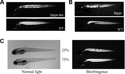
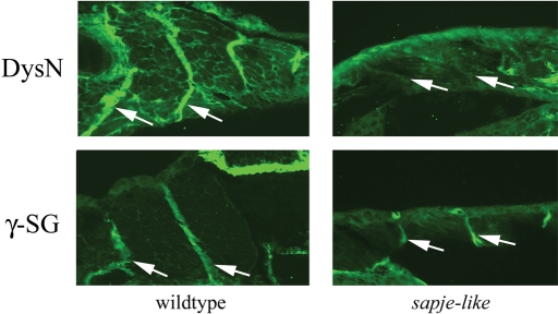
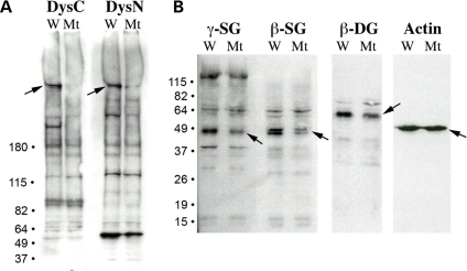
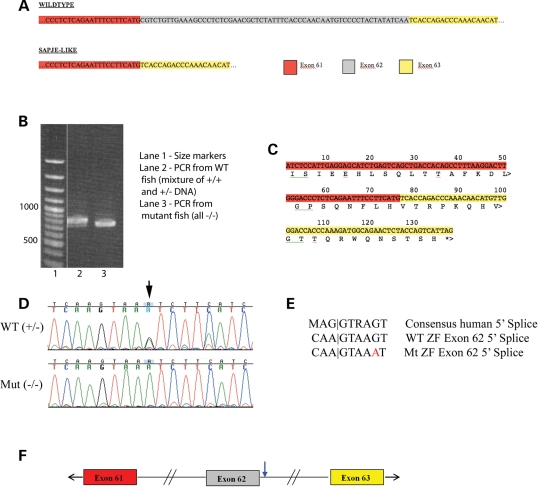
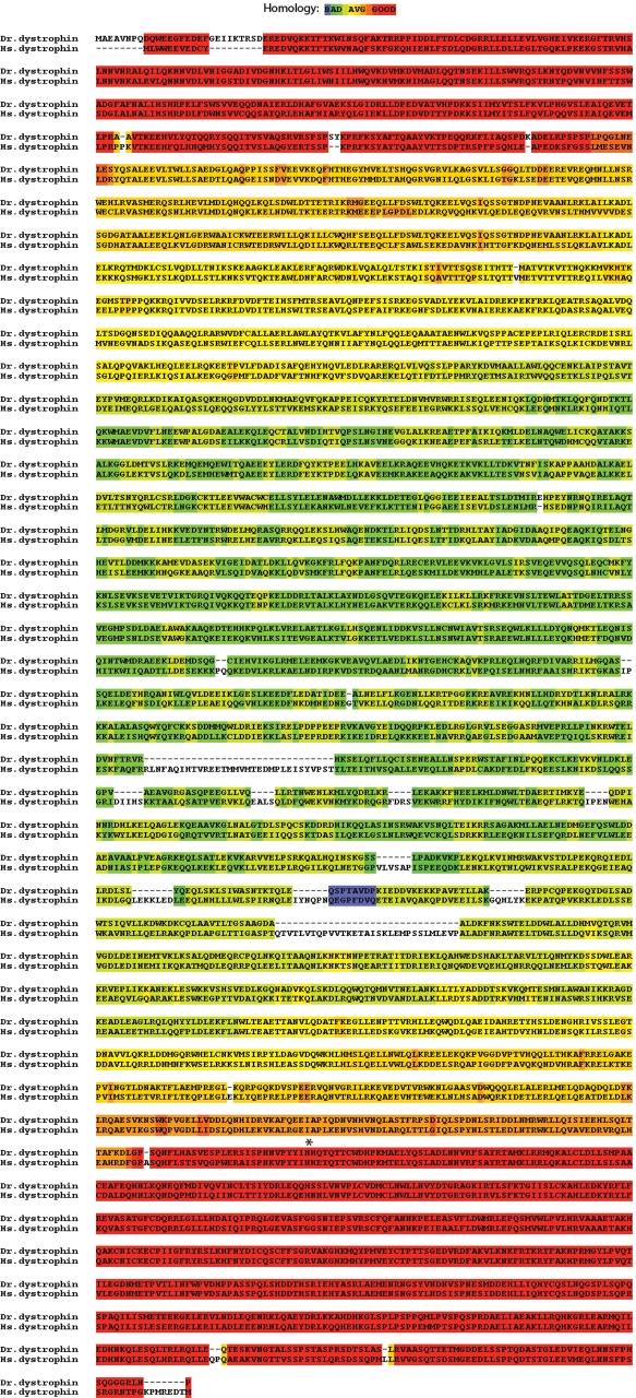
Sapje-like (sap(cl100)) was one of eight potential zebrafish muscle mutants isolated as part of an early-pressure screen of 500 families. This mutant shows a muscle tearing phenotype similar to sapje (dys-/-) and both mutants fail to genetically complement suggesting they have a mutation in the same gene. Protein analysis confirms a lack of dystrophin in developing sapje-like embryos. Sequence analysis of the sapje-like dystrophin mRNA shows that exon 62 is missing in the dystrophin transcript causing exon 63 to be translated out of frame terminating translation at a premature stop codon at the end of exon 63. Sequence analysis of sapje-like genomic DNA identified a mutation in the donor splice junction at the end of dystrophin exon 62. This mutation is similar to splicing mutations associated with human forms of Duchenne Muscular Dystrophy. Sapje-like is the first zebrafish dystrophin splicing mutant identified to date and represents a novel disease model which can be used in future studies to identify therapeutic compounds for treating diseases caused by splicing defects.


Figures

Figure 1.
Figure 1.
Sapje-like mutant offspring show muscle defects which can be assayed using birefringence. (A) Muscle degeneration in sapje-like mutants is clearly visible as small dark spots at 5 dpf using birefringence. These spots represent areas of muscle tearing. (B) The sapje-like birefringence is very similar to that seen for sapje, a known zebrafish dystrophin mutant. (C) Sapje and sapje-like fail to complement each other. Heterozygous sapje mutants (±) were crossed with heterozygous sapje-like mutants (±) and the offspring assayed at 4 dpf for defects in muscle using birefringence. From this cross, 25% of the offspring showed muscle lesions (top fish, right) suggesting that the two fish have mutations in the same gene. All panels are shown at approximately 10-fold magnification.
Figure 2.
Figure 2.
Immunohistochemical analysis of sapje-like horizontal sections at five days post-fertilization (dpf). Dystrophin protein localizes in unaffected siblings to the muscle myosepta (top left), but is absent in the mutant (top right). γ-Sarcoglycan is also strongly expressed in unaffected siblings (bottom left) and also in the sapje-like mutant (bottom right). From this analysis, it is difficult to determine if γ-sarcoglycan is down-regulated in the mutant. The myoseptas are designated with white arrows. For each antibody, pictures were taken at the same exposure level for both the wild-type and sapje-like mutant. All panels are shown at approximately 80-fold magnification.
Figure 3.
Figure 3.
Immunoblot analysis shows that dystrophin and the sarcoglycans are reduced in the sapje-like mutant. (A) Antibodies against both the C- and N-terminus of dystrophin (DysC and DysN, respectively) show that dystrophin is strongly down-regulated in the sapje-like mutant (‘Mt’) relative to the unaffected wild-type siblings (‘W’). (B) γ- and β-Sarcoglycan are down-regulated in the sapje-like mutant. β-Dystroglycan also appears to be affected in the sapje-like mutant, but the actin control and background non-specific bands seen in all of the blots show that the lanes were loaded equally. Size markers are shown on the left and each of the proteins is identified with an arrow on the blot.
Figure 4.
Figure 4.
Sapje-like has a mutation in the donor splice junction of dystrophin exon 62. (A) Based on sequencing the zebrafish dystrophin cDNA, zebrafish exon 61 is directly adjacent to exon 63 in the sapje-like mutant and missing exon 62. (B) Using primers within zebrafish dystrophin exons 57 and 63, PCR amplification of the zebrafish cDNA templates produces a smaller PCR product in the sapje-like mutant than in the unaffected siblings as assayed by agarose gel electrophoresis. (C) When exon 61 is directly adjacent to exon 63 as in sapje-like, this shifts the reading frame of exon 63 resulting in a premature stop codon at the end of exon 63. (D) Sequencing of the 5′ splice site adjacent to exon 62 reveals a G to A base change (see arrow). (E) Comparison of the human consensus 5′ splice site with the wild-type and mutant zebrafish dystrophin exon 62 5′ splice site. The sapje-like mutation resides at position +5 (G→A) and is highlighted in red. (F) The sapje-like mutation is located in the Exon 62 donor splice junction (blue arrow).
Figure 5.
Figure 5.
Conservation of the zebrafish dystrophin protein sequence. The zebrafish dystrophin sequence is designated ‘Dr. dystrophin’ and the human sequence is designated ‘Hs. Dystrophin’. The degree of homology has been evaluated by the Tcoffee alignment program (38,39) on a blue (not conserved) to red (highly conserved) scale. The location of the zebrafish sapje-like mutation is shown with an asterisk.

References

    1. Burghes A.H., Logan C., Hu X., Belfall B., Worton R.G., Ray P.N. A cDNA clone from the Duchenne/Becker muscular dystrophy gene. Nature. 1987;328:434–437. - PubMed
    1. Hoffman E.P., Brown R.H., Jr, Kunkel L.M. Dystrophin: the protein product of the Duchenne muscular dystrophy locus. Cell. 1987;51:919–928. - PubMed
    1. Monaco A.P., Neve R.L., Colletti-Feener C., Bertelson C.J., Kurnit D.M., Kunkel L.M. Isolation of candidate cDNAs for portions of the Duchenne muscular dystrophy gene. Nature. 1986;323:646–650. - PubMed
    1. Hoffman E.P., Fischbeck K.H., Brown R.H., Johnson M., Medori R., Loike J.D., Harris J.B., Waterston R., Brooke M., Specht L., et al. Characterization of dystrophin in muscle-biopsy specimens from patients with Duchenne's or Becker's muscular dystrophy. N. Engl. J. Med. 1988;318:1363–1368. - PubMed
    1. Flanigan K.M., von Niederhausern A., Dunn D.M., Alder J., Mendell J.R., Weiss R.B. Rapid direct sequence analysis of the dystrophin gene. Am. J. Hum. Genet. 2003;72:931–939. - PMC - PubMed

Publication types

MeSH terms