. 2008 Nov 15;121(Pt 22):3794-802.
doi: 10.1242/jcs.029678. Epub 2008 Oct 28.

Embryonic cardiomyocytes beat best on a matrix with heart-like elasticity: scar-like rigidity inhibits beating

Affiliations

Embryonic cardiomyocytes beat best on a matrix with heart-like elasticity: scar-like rigidity inhibits beating

Adam J Engler et al. J Cell Sci. .

Abstract

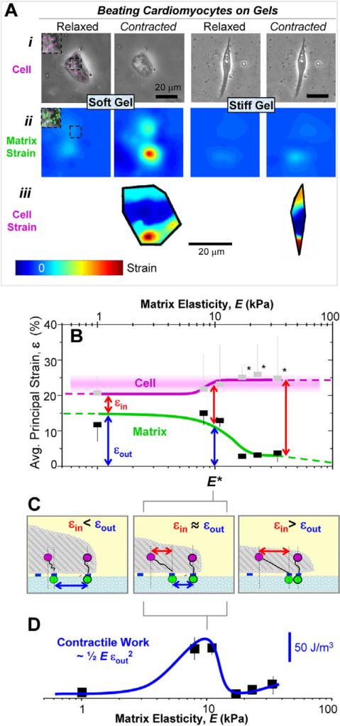
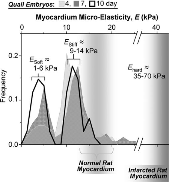
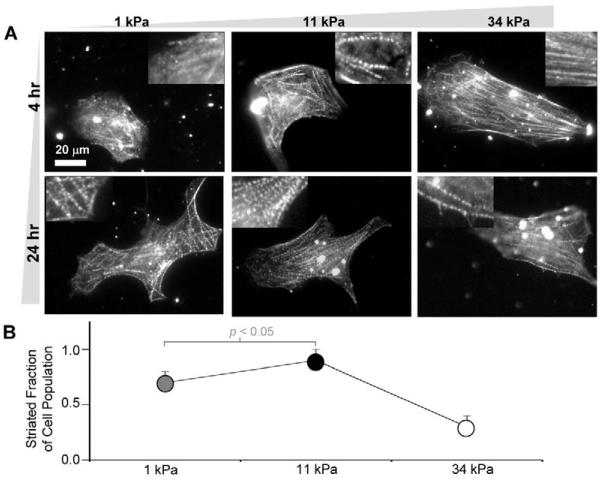
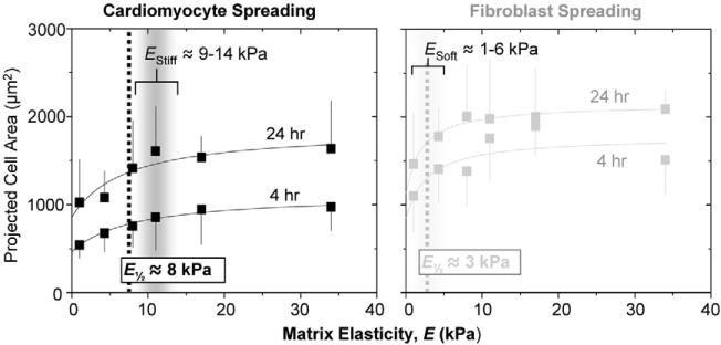
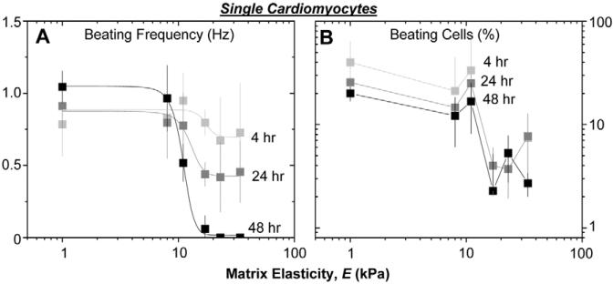
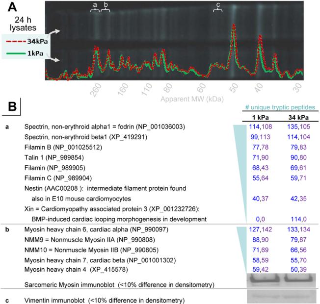
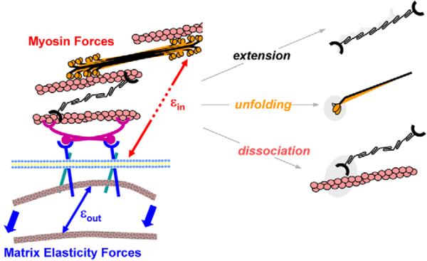
Fibrotic rigidification following a myocardial infarct is known to impair cardiac output, and it is also known that cardiomyocytes on rigid culture substrates show a progressive loss of rhythmic beating. Here, isolated embryonic cardiomyocytes cultured on a series of flexible substrates show that matrices that mimic the elasticity of the developing myocardial microenvironment are optimal for transmitting contractile work to the matrix and for promoting actomyosin striation and 1-Hz beating. On hard matrices that mechanically mimic a post-infarct fibrotic scar, cells overstrain themselves, lack striated myofibrils and stop beating; on very soft matrices, cells preserve contractile beating for days in culture but do very little work. Optimal matrix leads to a strain match between cell and matrix, and suggests dynamic differences in intracellular protein structures. A 'cysteine shotgun' method of labeling the in situ proteome reveals differences in assembly or conformation of several abundant cytoskeletal proteins, including vimentin, filamin and myosin. Combined with recent results, which show that stem cell differentiation is also highly sensitive to matrix elasticity, the methods and analyses might be useful in the culture and assessment of cardiogenesis of both embryonic stem cells and induced pluripotent stem cells. The results described here also highlight the need for greater attention to fibrosis and mechanical microenvironments in cell therapy and development.


Figures

Fig. 1
Fig. 1
Cell and matrix contraction. (A) Cells were imaged in brightfield mode 3−5 μm above the cell-matrix interface (i) and in fluorescence at the cell-matrix interface to observe particle motion during contraction (see inset pseudo-colored beads) to determine cellular and matrix deformations, respectively, that were then used to generate matrix (ii) and cellular (iii) principal strain maps. Note the difference in matrix strain between soft and stiff gels reflected by decreased deformation of the contracted cell. (B) The maximum cellular strain εcell is similar to the maximum matrix strain for soft matrices εout, resulting in small εin values. Hard substrates only allow the cell to deform itself during contraction. The transition between these regimes occurs at E*, where internal and external strain appear equivalent. Curve fits to guide the eye are of the form y=a+bEm×n/(Em+E1/2m) with m=6 and n=0 (matrix), n=1 (cell). Error bars, ± s.d. for at least five cells in duplicate studies. Paired t-tests were used to identify significant differences between εin of cells on softer versus stiffer gels (*P<0.05) and between cell strains on different gels. In the latter case, no two cell strains were significantly different. (C) Schematic of strained states for soft gels (E* gels) and stiff gels that respectively yield εinout, εinout, and εinout. (D) The contractile ‘work’ done by cardiomyocytes on the substrate peaks at E*.
Fig. 2
Fig. 2
Myocardial elasticity during embryogenesis. Histograms of elastic moduli (∼150 locations per sample) determined from quail embryos show two peaks, one indicating passive elasticity of contractile myocardium (EStiff) and a softer, second peak (ESoft) which surrounds the myocardium and increases in frequency with development. Results for normal and infarcted rat myocardium are indicated for comparison (Berry et al., 2006).
Fig. 3
Fig. 3
In vitro striation of cardiomyocytes. (A) Purified cardiomyocytes from 10-day-old embryonic myocardium were plated onto substrates of varying elasticity to observe striated cytoskeletal organization with skeletal α-actinin. Many cells on both soft gels and intermediate E* gels reassembled myofibrils whereas cells on hard matrices exhibited less myofibril reassembly. Inset images show magnified views of the larger images. (B) Fraction of cardiomyocytes that exhibit striation throughout the cytoplasm (± s.d. for >15 cells in triplicate studies). Organization appeared greatest on E* gels.
Fig. 4
Fig. 4
Cardiomyocyte and fibroblast spreading. Cardiomyocytes and pericardial fibroblasts were plated from 10-day old embryonic myocardium without purification and spread areas were measured (± s.d. for >15 cells in triplicate studies) after 4 or 24 hours. Fibroblasts were detected as α-actinin-negative cells and were the larger fraction of cells, consistent with in vivo proliferation (Fig. 2B). Half maximal ‘set points’ for projected cell area correspond closely to the peaks in passive tissue elasticity in Fig. 2B.
Fig. 5
Fig. 5
Cardiomyocyte beating is sensitive to matrix elasticity. Purified cardiomyocytes were plated at low density on matrices of varied stiffness, and beating was observed in phase contrast. (A) Beat frequency is elasticity-dependent, with cells on hard matrices slowing their beat frequency over days. (B) The percentage of cells beating was also quantified after 4, 24 and 48 hours post isolation. Beating cells persisted only on matrices with E≤E*. Error bars are ± s.d. for >5 cells, 10 seconds each in triplicate.
Fig. 6
Fig. 6
Protein abundance assessments in embryonic chicken cardiomyocytes grown on soft or hard matrices. (A) SDS-PAGE separation and densitometry analyses of lysates from 7-day embryonic chicken cardiomyocytes grown on 1 kPa or 34 kPa grown for 24 hours and then lysed, denatured and labeled by mBBr. (B) Mass spectrometry analyses of tryptic peptides or immunoblot results from the indicated gel bands (a-c). These bands were chosen as candidate cytoskeletal proteins using differential labeling in in-situ Cys-shotgun experiments (Table 1). The numbers of unique peptides are indicated from two different experiments. Immunoblotting for sarcomeric myosin and vimentin also indicated no significant differences in abundance.
Fig. 7
Fig. 7
Cell-driven, matrix-coupled remodeling through a combination of forced extension, unfolding or dissociation of proteins. On hard matrices, the intracellular strains εin exceed the extracellular strains εout and intracellular remodeling is promoted.

References

    1. Barry CP, Xie J, Lemmon V, Young AP. Molecular characterization of a multi-promoter gene encoding a chicken filamin protein. J. Biol. Chem. 1993;268:25577–25586. - PubMed
    1. Berry MF, Engler AJ, Woo YJ, Pirolli TJ, Bish LT, Jayasankar V, Morine KJ, Gardner TJ, Discher DE, Sweeney HL. Mesenchymal stem cell injection after myocardial infarction improves myocardial compliance. Am. J. Physiol. Heart Circ. Physiol. 2006;290:H2196–H2203. - PubMed
    1. Bird SD, Doevendans PA, van Rooijen MA, Brutel de la Riviere A, Hassink RJ, Passier R, Mummery CL. The human adult cardiomyocyte phenotype. Cardiovasc. Res. 2003;58:423–434. - PubMed
    1. Birks E, Hall J, Barton P, Grindle S, Latif N, Hardy J, Rider J, Banner N, Khaghani A, Miller L, et al. Gene profiling changes in cytoskeletal proteins during clinical recovery after left ventricular-assist device support. Circulation. 2005;112:157–164. - PubMed
    1. Breitbach M, Bostani T, Roell W, Xia Y, Dewald O, Nygren JM, Fries JW, Tiemann K, Bohlen H, Hescheler J, et al. Potential risks of bone marrow cell transplantation into infarcted hearts. Blood. 2007;110:1362–1369. - PubMed

Publication types