. 2008 Nov 4;105(44):16964-9.
doi: 10.1073/pnas.0809273105. Epub 2008 Oct 28.

Staged assembly of histone gene expression machinery at subnuclear foci in the abbreviated cell cycle of human embryonic stem cells

Affiliations

Staged assembly of histone gene expression machinery at subnuclear foci in the abbreviated cell cycle of human embryonic stem cells

Prachi N Ghule et al. Proc Natl Acad Sci U S A. .

Abstract

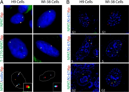
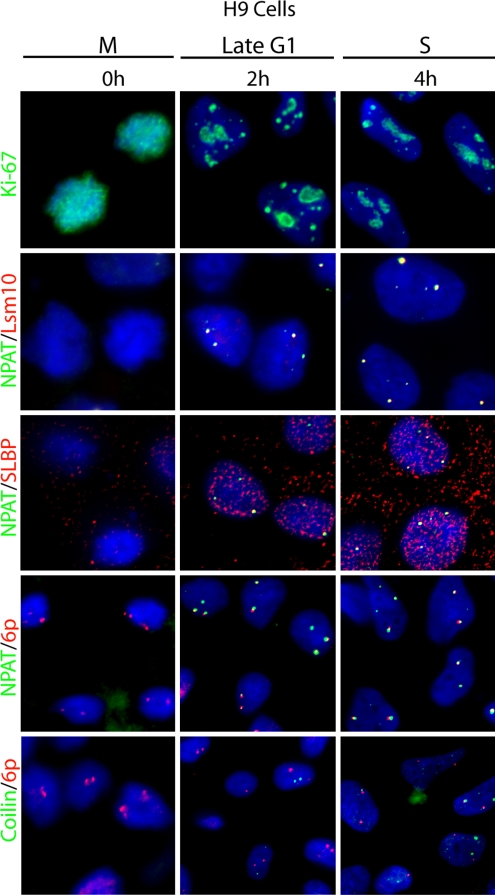
Human embryonic stem (hES) cells have an abbreviated G(1) phase of the cell cycle. How cells expedite G(1) events that are required for the initiation of S phase has not been resolved. One key regulatory pathway that controls G(1)/S-phase transition is the cyclin E/CDK2-dependent activation of the coactivator protein nuclear protein, ataxia-telangiectasia locus/histone nuclear factor-P (p220(NPAT)/HiNF-P) complex that induces histone gene transcription. In this study, we use the subnuclear organization of factors controlling histone gene expression to define mechanistic differences in the G(1) phase of hES and somatic cells using in situ immunofluorescence microscopy and fluorescence in situ hybridization (FISH). We show that histone gene expression is supported by the staged assembly and modification of a unique subnuclear structure that coordinates initiation and processing of transcripts originating from histone gene loci. Our results demonstrate that regulatory complexes that mediate transcriptional initiation (e.g., p220(NPAT)) and 3'-end processing (e.g., Lsm10, Lsm11, and SLBP) of histone gene transcripts colocalize at histone gene loci in dedicated subnuclear foci (histone locus bodies) that are distinct from Cajal bodies. Although appearance of CDK2-phosphorylated p220(NPAT) in these domains occurs at the time of S-phase entry, histone locus bodies are formed approximately 1 to 2 h before S phase in embryonic cells but 6 h before S phase in somatic cells. These temporal differences in the formation of histone locus bodies suggest that the G(1) phase of the cell cycle in hES cells is abbreviated in part by contraction of late G(1).


Conflict of interest statement

The authors declare no conflict of interest.

Figures

Fig. 1.
Fig. 1.
The p220NPAT foci are associated with histone gene loci in H9 hES cells and normal diploid WI-38 fibroblasts. (A) IF microscopy images were obtained using antibodies against p220NPAT (green), phospho-T1270-p220NPAT (green), or coilin (blue), and a DIG-labeled FISH probe adjacent to the histone gene cluster on 6p22 (red). The arrows in the bottom row, left column, indicate the position of coilin foci. The Insets in the lower right of each panel in the bottom row indicate colocalization between p220NPAT/colin/6p. DAPI staining (blue) is used to visualize the nucleus (top 2 rows). There are typically 2 or 4 p220NPAT foci, depending on the cell cycle stage, that are consistently in proximity to histone gene clusters. In 50–60% of cells, coilin foci (Cajal bodies) overlap with at least one p220NPAT foci. (B) IF images as in A, using antibodies against p220NPAT (green) and Ki-67 (blue), a marker for cell cycle position, and the 6p22 FISH probe. Cell cycle stages are indicated at the lower left of each panel.
Fig. 2.
Fig. 2.
Colocalization of factors mediating histone gene transcriptional initiation and 3′-end transcript processing of histone mRNA. IF microscopy images were obtained for H9 hES cells (Left column) and normal diploid WI-38 cells (Right column) using antibodies against p220NPAT (green) and factors that process or interact with histone transcripts (Lsm10, Lsm11, SLBP, or 3′ hExo; red). SLBP interacts with the 3′ hairpin in histone mRNA; the protein only partially colocalizes with p220NPAT foci. Foci of 3′ hExo show no colocalization with p220NPAT foci (green, row 4) and complete overlap with PML/ND10 bodies (green, row 5) in both hES cells and somatic WI-38 cells. The percentages in the lower left of the panels represent positive cells for colocalization of respective factors in each cell type.
Fig. 3.
Fig. 3.
Association of p220NPAT and 3′-end processing factors with histone gene loci during the hES cell cycle. Mitotically synchronized hES cells at various cell cycle stages were monitored by IF microscopy for association of Lsm10 or SLBP (red) with p220NPAT (green; rows 2 and 3), and spatial linkage of the histone gene cluster at 6p22 (red) with p220NPAT or coilin (green; rows 4 and 5). Ki-67 (green) staining (row 1) was done to establish cell cycle position, and DAPI staining (all rows; blue) was used to visualize the nucleus.
Fig. 4.
Fig. 4.
Colocalization of 3′-end processing factors SLBP and Lsm10 with p220NPAT during the cell cycle in somatic cells. Synchronized WI-38 fibroblasts were examined at various cell cycle stages by IF microscopy for spatial interactions between Lsm10 or SLBP (red) with p220NPAT (green). Ki-67 (green) staining (Top row) was done to establish cell cycle position, and DAPI staining (all rows; blue) was used to visualize the nucleus. The percentages in the lower left of the panels represent cells positive for Ki-67 (Top row) and SLBP (Bottom row). The images in row 1 were taken at 40× magnification.

References

    1. Becker KA, et al. Self-renewal of human embryonic stem cells is supported by a shortened G1 cell cycle phase. J Cell Physiol. 2006;209:883–893. - PubMed
    1. Becker KA, Stein JL, Lian JB, van Wijnen AJ, Stein GS. Establishment of histone gene regulation and cell cycle checkpoint control in human embryonic stem cells. J Cell Physiol. 2007;210:517–526. - PubMed
    1. Pardee AB. A restriction point for control of normal animal cell proliferation. Proc Natl Acad Sci USA. 1974;71:1286–1290. - PMC - PubMed
    1. Pardee AB. G1 events and regulation of cell proliferation. Science. 1989;246:603–608. - PubMed
    1. Stein GS, Stein JL, van Wijnen AJ, Lian JB. Transcriptional control of cell cycle progression: The histone gene is a paradigm for the G1/S phase and proliferation/differentiation transitions. Cell Biol Int. 1996;20:41–49. - PubMed

Publication types

MeSH terms

LinkOut - more resources