. 2008 Nov 4;105(44):16928-33.
doi: 10.1073/pnas.0808427105. Epub 2008 Oct 28.

Urea denaturation by stronger dispersion interactions with proteins than water implies a 2-stage unfolding

Affiliations

Urea denaturation by stronger dispersion interactions with proteins than water implies a 2-stage unfolding

Lan Hua et al. Proc Natl Acad Sci U S A. .

Abstract

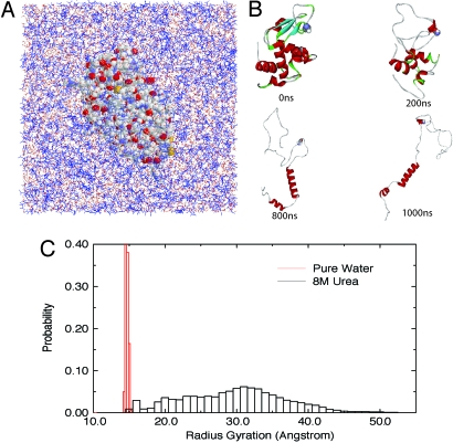
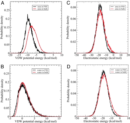
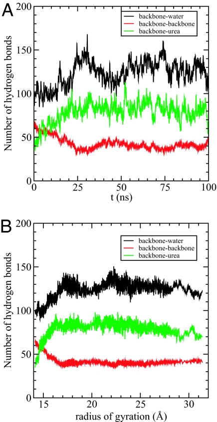
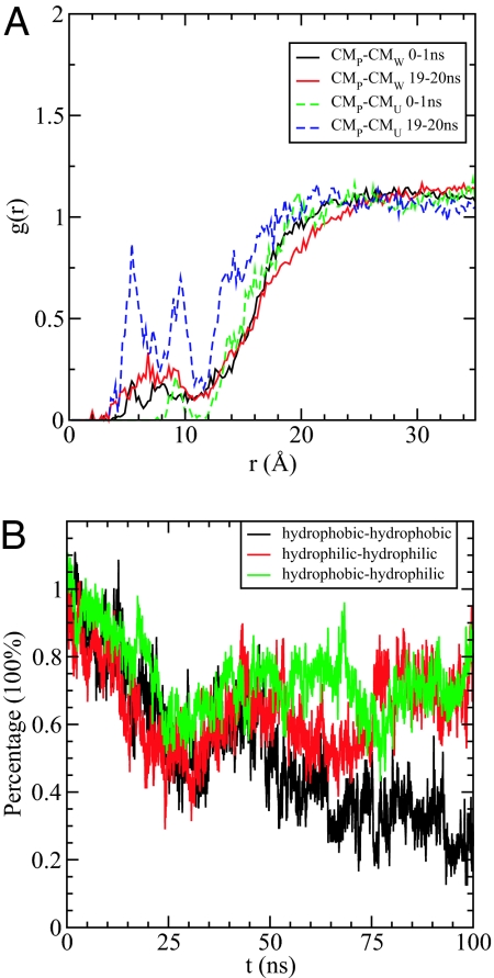
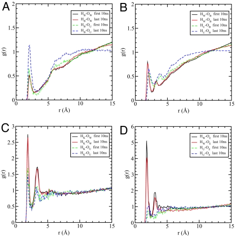
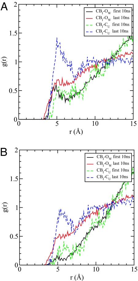
The mechanism of denaturation of proteins by urea is explored by using all-atom microseconds molecular dynamics simulations of hen lysozyme generated on BlueGene/L. Accumulation of urea around lysozyme shows that water molecules are expelled from the first hydration shell of the protein. We observe a 2-stage penetration of the protein, with urea penetrating the hydrophobic core before water, forming a "dry globule." The direct dispersion interaction between urea and the protein backbone and side chains is stronger than for water, which gives rise to the intrusion of urea into the protein interior and to urea's preferential binding to all regions of the protein. This is augmented by preferential hydrogen bond formation between the urea carbonyl and the backbone amides that contributes to the breaking of intrabackbone hydrogen bonds. Our study supports the "direct interaction mechanism" whereby urea has a stronger dispersion interaction with protein than water.


Conflict of interest statement

The authors declare no conflict of interest.

Figures

Fig. 1.
Fig. 1.
Unfolding of lysozyme in 8 M urea. (A) The solvated system of W62G mutant lysozyme in 8 M urea (the protein is represented by VDW balls, and solvent by wires with urea colored blue). (B) A few snapshots of the mutant lysozyme during 1-μs unfolding simulation (at 0 ns, 200 ns, 800 ns, and 1,000 ns). (C) Comparison of the radius gyration of protein lysozyme in pure water and 8 M urea. (The 8 M urea data are from an aggregate of 5-μs simulations, and the pure water data are from 100-ns simulation only because the protein is fairly stable in pure water at room temperature. See text for more details.)
Fig. 2.
Fig. 2.
Interaction energy distribution. (A and B) The probability distribution function of Van der Waals energy of urea (A) and water (B) in the first solvation shell of mutant lysozyme and in the bulk region, respectively, with the rest of system. (C and D) The probability distribution function of electrostatic energy of urea (C) and water (D) in the first solvation shell of mutant lysozyme and in the bulk region, respectively, with the rest of system. The first solvation shell (FSS) is defined as within 5.0 Å of protein and the bulk region is defined as not within 6.0 Å of protein.
Fig. 3.
Fig. 3.
Hydrogen bonds with backbone. (A) The number of hydrogen bonds formed between backbone and water (black), between backbone and backbone (red), and between backbone and urea (green), respectively, as a function of time. (B) The number of hydrogen bonds as a function of the radius of gyration of the mutant lysozyme. Black is for backbone–water hydrogen bonds, red is for backbone–backbone hydrogen bonds, and green is for backbone–urea hydrogen bonds.
Fig. 4.
Fig. 4.
Dry globule formation. (A) The radial distribution functions g(r) between the center of mass (COM) of the protein and the COM of urea and water in the first 1 ns and 19th ns (19–20 ns). It clearly shows that the concentration of urea increases much faster than water within the core of the protein (≈20 Å), indicating that urea might intrude into the protein ahead of water. (B) The time dependence of the percentage of the initial backbone–backbone hydrogen bonds formed by the residue pairs of hydrophobic–hydrophobic (black), hydrophilic–hydrophilic (red), and hydrophobic–hydrophilic (green), respectively.
Fig. 5.
Fig. 5.
Radial distribution functions. (A) The pair radial distribution function g(r) between backbone amide hydrogen HB and water oxygen OW (solid), as well as urea oxygen OU (dash). (B) The pair radial distribution function g(r) between backbone carbonyl oxygen OB and water hydrogen HW (solid), as well as urea hydrogen HU (dash). (C) The pair radial distribution function g(r) between positively charged lysine side-chain hydrogen HK and water oxygen OW (solid), as well as urea oxygen OU (dash). (D) The pair radial distribution function g(r) between negatively charged glutamic acid side-chain oxygen OE and water hydrogen HW (solid), as well as urea hydrogen HU (dash). All of the g(r) functions are averaged over the first 10 ns (black, green) and the last 10 ns (red, blue) of total 100 ns, respectively.
Fig. 6.
Fig. 6.
Enhanced solubility of nonpolar residues. (A) The pair radial distribution function g(r) between the β-carbon of isoleucine CBI and center of mass of solvent, represented by oxygen atom of water OW and carbon atom of urea CU. (B) The same as A, except that urea or water that forms hydrogen bonds with backbone is excluded in the calculation of g(r). All of the g(r) functions are averaged over the first 10 ns (black, green) and the last 10 ns (red, blue) of total 100 ns, respectively.

Comment in

  • Protein denaturation by urea: slash and bond.
    Rossky PJ. Rossky PJ. Proc Natl Acad Sci U S A. 2008 Nov 4;105(44):16825-6. doi: 10.1073/pnas.0809224105. Epub 2008 Oct 30. Proc Natl Acad Sci U S A. 2008. PMID: 18974225 Free PMC article. No abstract available.

References

    1. Pace C. Determination and analysis of urea and guanidine hydrochloride denaturation curves. Methods Enzymol. 1986;131:266–280. - PubMed
    1. Schellman J. The stability of hydrogen-bonded peptide structures in aqueous solution. Trav Lab Carlsberg Ser Chim. 1955;29:230–259. - PubMed
    1. Tanford C. Protein denaturation. C. Theoretical models for the mechanism of denaturation. Adv Protein Chem. 1970;24:1–95. - PubMed
    1. Alonso D, Dill K. Solvent denaturation and stabilization of globular proteins. Biochemistry. 1991;30:5974–5985. - PubMed
    1. Scholtz J, Barrick D, York E, Stewart J, Baldwin R. Urea unfolding of peptide helices as a model for interpreting protein unfolding. Proc Natl Acad Sci USA. 1995;92:185–189. - PMC - PubMed

Publication types

LinkOut - more resources