. 2008;3(10):e3511.
doi: 10.1371/journal.pone.0003511. Epub 2008 Oct 29.

Remote excitation of neuronal circuits using low-intensity, low-frequency ultrasound

Affiliations

Remote excitation of neuronal circuits using low-intensity, low-frequency ultrasound

William J Tyler et al. PLoS One. 2008.

Abstract

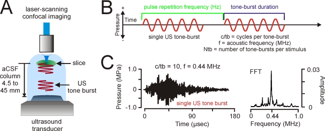
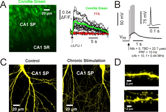
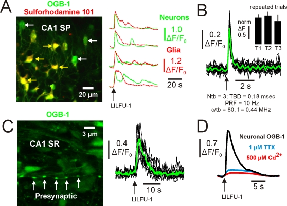
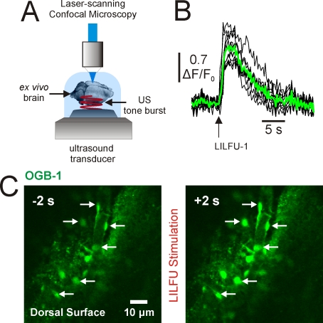
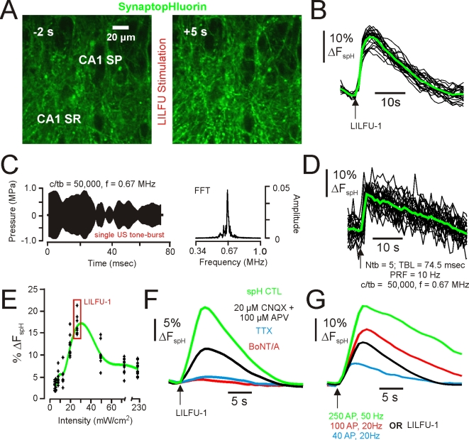
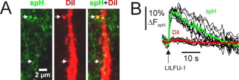
Possessing the ability to noninvasively elicit brain circuit activity yields immense experimental and therapeutic power. Most currently employed neurostimulation methods rely on the somewhat invasive use of stimulating electrodes or photon-emitting devices. Due to its ability to noninvasively propagate through bone and other tissues in a focused manner, the implementation of ultrasound (US) represents a compelling alternative approach to current neuromodulation strategies. Here, we investigated the influence of low-intensity, low-frequency ultrasound (LILFU) on neuronal activity. By transmitting US waveforms through hippocampal slice cultures and ex vivo mouse brains, we determined LILFU is capable of remotely and noninvasively exciting neurons and network activity. Our results illustrate that LILFU can stimulate electrical activity in neurons by activating voltage-gated sodium channels, as well as voltage-gated calcium channels. The LILFU-induced changes in neuronal activity were sufficient to trigger SNARE-mediated exocytosis and synaptic transmission in hippocampal circuits. Because LILFU can stimulate electrical activity and calcium signaling in neurons as well as central synaptic transmission we conclude US provides a powerful tool for remotely modulating brain circuit activity.


Conflict of interest statement

Competing Interests: The authors have declared that no competing interests exist.

Figures

Figure 1
Figure 1. Generation and propagation of LILFU waveforms through neuronal tissue.
(A) General experimental configuration implemented to transmit LILFU waveforms through slice cultures while optically monitoring neuronal activity. (B) Graphical illustration of some of the variables involved in constructing LILFU waveforms. These variables include acoustic frequency (f), the number of acoustic cycles per tone burst (c/tb), tone burst duration (TBD), pulse repetition frequency (PRF), and number of tone bursts per stimulus (Ntb). (C) Acoustic pressure wave (left) produced by a typical US tone burst consisting of 10 acoustic cycles at f = 0.44 MHz and FFT of this US tone burst (right). For the construction of our primary US stimulus waveform (LILFU-1), we used a linearly sweeping PRF by repeating the illustrated tone burst from 0–100 Hz over a 5 sec period.
Figure 2
Figure 2. LILFU stimulates sodium transients mediated by voltage-gated sodium channels in hippocampal neurons.
(A) Confocal image (left) of a slice culture loaded with CoroNa Green AM. Hippocampal regions CA1 stratum pyramidale (SP) and stratum radiatum (SR) are illustrated. Individual (black) and averaged (color) Na+ transients (right) triggered in CA1 pyramidal neuron somas by LILFU-1 under control conditions and in the presence of TTX. (B) Voltage trace of membrane voltage in response to five US tone bursts delivered at a PRF of 10 Hz during whole-cell current clamp recordings of a CA1 pyramidal neuron. (C) Neuronal membrane integrity is preserved following chronic in vitro stimulation with LILFU. Confocal images of CA1 pyramidal neurons from hippocampal slice cultures prepared from thy-1-YFP mice. The images shown are from a control slice culture (left) and a slice culture following chronic stimulation (right) with LILFU-1 every 8 min for 48 h (360 LILFU-1 stimuli). (D) Similar to (C), but higher magnification images of regions in CA1 SR, which more clearly illustrate the presence of fine membrane structures such as dendritic spines for control (top) and chronic LILFU stimulation conditions (bottom).
Figure 3
Figure 3. LILFU triggers voltage-dependent somatic and presynaptic Ca2+ transients in neurons.
(A) Confocal image (left) of a slice culture loaded with OGB-1 AM (green) to monitor Ca2+ activity and Sulforhodamine 101 (red) to identify glial cells (yellow). Representative LILFU-triggered Ca2+ transients observed in the somas of neurons and glial cells are illustrated (right). (B) Individual (black) and averaged (green) Ca2+ transients observed in the somas of neurons in response to a brief LILFU waveform. The histogram (inset) illustrates trial 1 normalized mean Ca2+ transient amplitudes in response to repeated trials of LILFU stimulation (n = 19 cells from 3 slices). (C) Confocal image (left) of a slice culture loaded with OGB-1 AM illustrating en passant boutons located in CA1 SR. Individual (black) and averaged (green) presynaptic Ca2+ transients (right) produced by stimulation with LILFU-1. (D) Averaged somatic Ca2+ transients obtained from neurons under control conditions or in the presence of either TTX (n = 36 from 4 slices) or Cd2+ (n = 30 from 4 slices) in response to stimulation with LILFU-1.
Figure 4
Figure 4. LILFU waveforms transmitted through whole brains are capable of stimulating calcium transients.
(A) Illustration of basic experimental procedure we developed to transmit LILFU waveforms through whole ex vivo brains prepared from adult wild-type mice and bath-loaded with OGB-1 AM. As depicted, LILFU waveforms were transmitted from the ventral surface of the brain through the tissue to the dorsal surface where we performed confocal imaging. (B) Individual (black) and averaged (green) Ca2+ transients observed in the somas of cells on the dorsal surface of an ex vivo brain in response to stimulation with LILFU-1, which was transmitted through the brain from the ventral surface. (C) Confocal images illustrating OGB-1 loaded cells on the dorsal surface of the brain. The image on left illustrates cells during baseline, while the image on the right illustrates cells two-seconds after stimulation with LILFU-1 ensued.
Figure 5
Figure 5. LILFU stimulates SNARE-mediated synaptic vesicle exocytosis and central synaptic transmission.
(A) Confocal images illustrating spH signals obtained before (left) and during (right) stimulation with LILFU-1. (B) Individual (black) and averaged (green) spH signals typically obtained in response to stimulation with LILFU-1. (C) Acoustic pressure wave (left) produced by a single LILFU tone burst consisting of 50,000 acoustic cycles at f = 0.67 MHz and FFT of LILFU tone burst (right). (D) Individual (black) and averaged (green) spH signals obtained in response to stimulation with the LILFU tone burst shown in (C) delivered at a PRF = 10 Hz for 0.5 s to produce Np = 5. (E) Histogram of spH responses obtained as a function of acoustic intensity. Responses from individual experiments are indicated by black crosses while the average response is indicated by the green line. (F) Averaged spH signals illustrating the effect of CNQX+APV (n = 84 from 4 slices), TTX (n = 108 from 4 slices), or BoNT/A (n = 60 from 4 slices) on synaptic vesicle exocytosis induced by LILFU-1. (G) Averaged spH signals obtained from buttons in response to field stimulation of Schaffer collaterals with 250 AP, 50 Hz (n = 48), 100 AP, 20 Hz (n = 63), 40 AP, 20 Hz (n = 51), or by LILFU-1 (n = 148).
Figure 6
Figure 6. Influence of LILFU on putative excitatory hippocampal CA3-CA1 synapses.
(A) Confocal images illustrating spH expression in CA1 SR (left) and an apical dendritic branch of a CA1 pyramidal neuron, which was labeled with DiI using a DiOlistic labeling technique (middle). The two-channel confocal image (right) illustrates putative excitatory synapses indicated by apposition of spH+ puncta and dendritic spines. (B) Individual (black), mean spH (green), and mean DiI (red) signals obtained from terminals impinging on dendritic spines in response to stimulation with LILFU-1.

References

    1. Wagner T, Valero-Cabre A, Pascual-Leone A. Noninvasive Human Brain Stimulation. Annu Rev Biomed Eng. 2007;9:527–565. - PubMed
    1. Zhang F, Wang LP, Brauner M, Liewald JF, Kay K, et al. Multimodal fast optical interrogation of neural circuitry. Nature. 2007;446:633–639. - PubMed
    1. Gavrilov LR, Gersuni GV, Ilyinsky OB, Sirotyuk MG, Tsirulnikov EM, et al. The effect of focused ultrasound on the skin and deep nerve structures of man and animal. Prog Brain Res. 1976;43:279–292. - PubMed
    1. Fry FJ, Ades HW, Fry WJ. Production of reversible changes in the central nervous system by ultrasound. Science. 1958;127:83–84. - PubMed
    1. ter Haar G. Therapeutic applications of ultrasound. Prog Biophys Mol Biol. 2007;93:111–129. - PubMed

Publication types