. 2008 Aug 25:2:8.
doi: 10.3389/neuro.09.008.2008. eCollection 2008.

Feeling present in arousing virtual reality worlds: prefrontal brain regions differentially orchestrate presence experience in adults and children

Affiliations

Feeling present in arousing virtual reality worlds: prefrontal brain regions differentially orchestrate presence experience in adults and children

Thomas Baumgartner et al. Front Hum Neurosci. .

Abstract

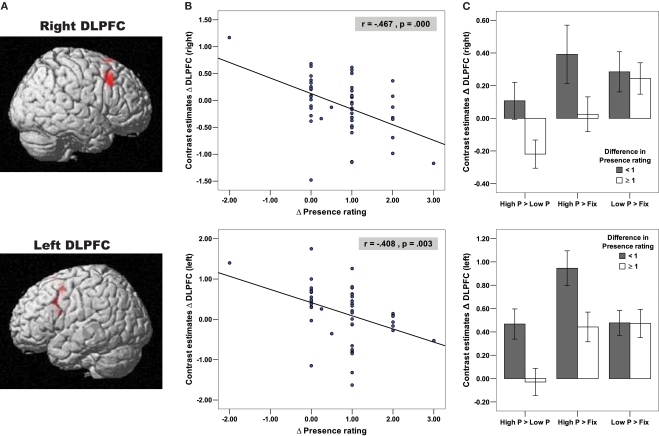
Virtual reality (VR) is a powerful tool for simulating aspects of the real world. The success of VR is thought to depend on its ability to evoke a sense of "being there", that is, the feeling of "Presence". In view of the rapid progress in the development of increasingly more sophisticated virtual environments (VE), the importance of understanding the neural underpinnings of presence is growing. To date however, the neural correlates of this phenomenon have received very scant attention. An fMRI-based study with 52 adults and 25 children was therefore conducted using a highly immersive VE. The experience of presence in adult subjects was found to be modulated by two major strategies involving two homologous prefrontal brain structures. Whereas the right DLPFC controlled the sense of presence by down-regulating the activation in the egocentric dorsal visual processing stream, the left DLPFC up-regulated widespread areas of the medial prefrontal cortex known to be involved in self-reflective and stimulus-independent thoughts. In contrast, there was no evidence of these two strategies in children. In fact, anatomical analyses showed that these two prefrontal areas have not yet reached full maturity in children. Taken together, this study presents the first findings that show activation of a highly specific neural network orchestrating the experience of presence in adult subjects, and that the absence of activity in this neural network might contribute to the generally increased susceptibility of children for the experience of presence in VEs.

Keywords: adults; brain maturation; children; cognitive/executive control; prefrontal cortex; presence experience; virtual reality.


Figures

Figure 1
Figure 1
Experimental design and Presence rating. (A) Experimental paradigm. Different roller coaster scenarios were presented by means of MR-compatible goggles and earphones. Whereas in the High Presence condition the roller coaster ride consisted of spectacular ascending and descending sections and loops, in the Low Presence condition the roller coaster cart followed a winding but horizontal path. (B) Depicted is the mean ± S.E.M. in Presence rating on a 5-point scale, broken down for group (adults/children) and condition (High Presence/Low Presence). The scale measures the subjective impression of how much the subjects felt they were situated in the midst of the action of the roller coaster ride rather than merely observing it. Both adults and children clearly indicated an enhanced presence experience in the High compared to the Low Presence condition. (C) Similar distribution of presence ratings in adults and children, indicating that about 40% of subjects in each group reported a difference in presence rating of less than one between the High and Low Presence condition (referred to in the paper as low Presence rating group), whereas about 60% of subjects in each group reported a difference in the presence rating of higher than or equal to one (referred to as high Presence rating group).
Figure 2
Figure 2
Negative correlation of bilateral DLPFC with Presence experience in adults. (A) Depicted on the brain images are the bilateral DLPFC (right: x = 48, y = 21, z = 39; BA = 9; left: x = −54, y = 15, z = 36, BA = 9), which negatively correlated with the presence rating in adult subjects, indicating that the High Presence condition yielded a greater activation increase in the bilateral DLPFC in adult subjects who reported a smaller amount of Presence elevation between the High and Low Presence condition. (B) These negative correlations of DLPFC and presence ratings are depicted on the two scatter plots using functional ROIs. (C) In order to examine whether DLPFC activation differences in the High Presence (as expected), Low Presence or in both conditions contributed to the negative correlational pattern, we extracted contrast estimates difference in the bilateral DLPFC for the contrasts Low P > Fix, High P > Fix and High P > Low P (High P = High Presence, Low P = Low Presence and Fix = Fixation Baseline). Based on these contrast estimates, we created bar plots, broken down for adult subjects with difference in Presence rating < or ≥1, referred to as low and high Presence rating group, respectively. These bar plots illustrated that there is no difference in DLPFC activation between the two rating groups during the Low Presence condition (independent t-tests for right DLPFC: t = 0.26, df = 50, p = 0.793 and left DLPFC: t = 0.02, df = 50, p = 0.977). In contrast, during the High Presence condition, the low Presence rating group showed an increase, while the high Presence rating group showed no change (left DLPFC) or even a decrease in DLPFC (right DLPFC) activation, resulting in a significant group difference in the High Presence condition (independent t-tests for right DLPFC (one-tailed): t = 1.87, df = 50, p = 0.033 and left DLPFC: t = 2.54, df = 50, p = 0.014). The rating groups significantly differed therefore in the contrast High P > Low P for the right (independent t-test: t = 2.32, df = 50, p = 0.024) and the left DLPFC (independent t-test: t = 2.80, df = 50, p = 0.007).
Figure 3
Figure 3
Negative connectivity with right and positive connectivity with left DLPFC in adults. (A) Negative connectivity (blue colour) with right DLPFC in the dorsal visual stream (including superior and inferior parietal gyrus as well as superior occipital gyrus) and sensory-motor areas in adult subjects (at p < 0.005, cluster extent: 10 voxels; SPG, superior parietal gyrus; IPG, inferior parietal gyrus; MOG, middle occipital gyrus; SOG, superior occipital gyrus). (B) Significant differences in negative connectivity between right DLPFC and left DLPFC in areas depicted in (A), at p < 0.005 (yellow), p < 0.01 (violet) and p < 0.05 (green, all with a cluster extent of 10 voxels). Most areas, except for the ones in blue colour, depict a clear right-sided lateralization pattern in negative connectivity. (C) Positive connectivity (red colour) with left DLPFC in medial PFC (including ACC), extrastriate visual cortex and subcortical areas (including dorso-medial Thalamus and Brainstem) in adult subjects (at p < 0.005, cluster extent: 10 voxels; MPFC, medial prefrontal cortex; Bst, Brainstem; Tha, Thalamus; Cau, Caudatus; PHiG, Parahippocampal Gyrus). (D) Significant differences in positive connectivity between left DLPFC and right DLPFC in areas depicted in (C), at p < 0.005 (yellow), p < 0.01 (violet) and p < 0.05 (green, all with a cluster extent of 10 voxels, areas in red colour illustrate no significant difference). All areas, except for a few voxels in the medial PFC in red colour, depict a clear left-sided lateralization pattern in positive connectivity in adult subjects.
Figure 4
Figure 4
Positive connectivity with right DLPFC in children. (A) Positive connectivity (red colour) with right DLPFC mainly in subcortical and emotional areas (including amygdala/hippocampus and insula) as well as multisensory integration areas (posterior STG), areas of the ventral visual processing stream (including Fusiform Gyrus) and small clusters in prefrontal areas (inferior and medial PFC) in children subjects (at p < 0.005, cluster extent: 10 voxels; Amy, amygdala; Hip, hippocampus; ITG/MTG,STG, inferior/middle/superior temporal gyrus; Put, putamen; IFG, inferior frontal gyrus; MPFC, medial prefrontal cortex). (B) Significant differences in positive connectivity between right DLPFC and left DLPFC in areas depicted in (A), at p < 0.005 (yellow), p < 0.01 (violet) and p < 0.05 (green, all with a cluster extent of 10 voxels). In children subjects, all areas depict a clear right-sided lateralization pattern in positive connectivity.
Figure 5
Figure 5
Negative connectivity differences in adults: Low Presence rating group > High Presence rating group. (A) Whereas all adults, irrespective of their presence rating, used their right DLPFC to down-regulate activation in the dorsal visual stream and sensory-motor areas (depicted in dark blue colour, the same activation as in Figure 3A), subjects of the low Presence rating group used their right DLPFC to down-regulate additional areas of this dorsal visual system (including bilateral precuneus, inferior and superior parietal gyrus) as well as the posterior thalamus (depicted in light blue colour; p < 0.005, cluster extent: 10 voxels; PreCu, Precuneus; IPG, inferior parietal gyrus; SPG, superior parietal gyrus; PCC, posterior cingulate cortex; Tha, Thalamus). Areas in violet colour are down-regulated by all adult subjects, but subjects of the low Presence rating group showed an even stronger down-regulation in this part of the inferior and superior parietal cortex. For those regions showing a differential group effect depicted in (A), we also created regions of interests and extracted Beta estimates. Bar plots based on these Beta estimates are depicted for (B) adults and (C) children, broken down for the low (Difference in Presence rating < 1) and high (Difference in Presence rating ≥ 1) Presence rating groups. Positive values indicate negative connectivity, whereas negative values indicate positive connectivity. Asterisk indicate significant increase in negative or positive connectivity at p < 0.05 (*), p ≤ 0.01 (**), p ≤ 0.005 (***), or p ≤ 0.001 (****). The bar plots in (B) illustrate that, except for the thalamus, an unilateral, right-sided and negative connectivity pattern has been observed in adult subjects of the low Presence rating group. In contrast, children of the low Presence rating group did not show this negative connectivity pattern in any brain region of the dorsal visual stream, either with the right or left DLPFC [depicted in (C), see Table S12 for detailed statistical analyses].

References

    1. Amodio D. M., Frith C. D. (2006). Meeting of the minds: the medial frontal cortex and social cognition. Nat. Rev. Neurosci. 7, 269–27710.1038/nrn1884 - DOI - PubMed
    1. Annett M. (1970). A classification of hand preference by association analysis. Br. J. Clin. Psychol. 61, 303–321 - PubMed
    1. Aron A. R., Robbins T. W., Poldrack R. A. (2004). Inhibition and the right inferior frontal cortex. Trends Cogn. Sci. 8(4), 170–17710.1016/j.tics.2004.02.010 - DOI - PubMed
    1. Ashburner J., Friston K. J. (2000). Voxel-based morphometry – the methods. Neuroimage 11(6 Pt 1), 805–82110.1006/nimg.2000.0582 - DOI - PubMed
    1. Ashburner J., Friston K. J. (2005). Unified segmentation. Neuroimage 26(3), 839–85110.1016/j.neuroimage.2005.02.018 - DOI - PubMed