. 2008 Mar 28:1:5.
doi: 10.3389/neuro.09.005.2007. eCollection 2007.

Neural substrate of concurrent sound perception: direct electrophysiological recordings from human auditory cortex

Affiliations

Neural substrate of concurrent sound perception: direct electrophysiological recordings from human auditory cortex

Aurélie Bidet-Caulet et al. Front Hum Neurosci. .

Abstract

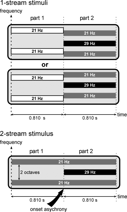
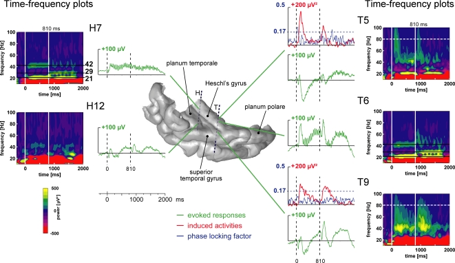
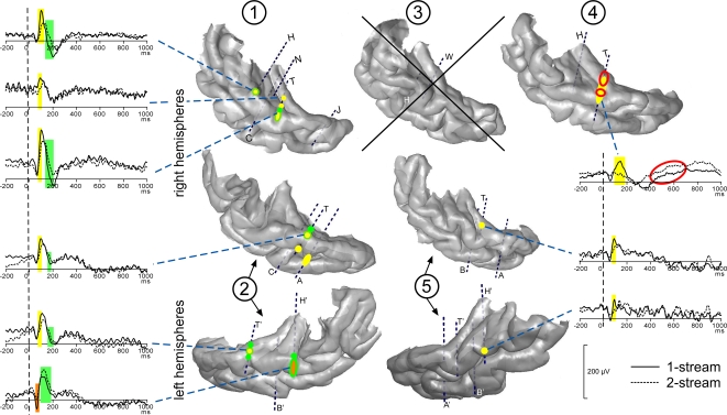
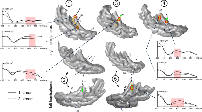
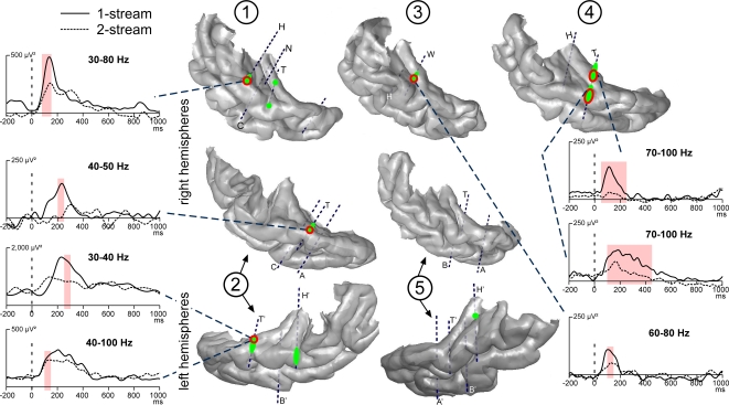
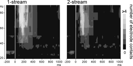
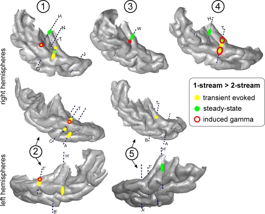
In everyday life, consciously or not, we are constantly disentangling the multiple auditory sources contributing to our acoustical environment. To better understand the neural mechanisms involved in concurrent sound processing, we manipulated sound onset asynchrony to induce the segregation or grouping of two concurrent sounds. Each sound consisted of amplitude-modulated tones at different carrier and modulation frequencies, allowing a cortical tagging of each sound. Electrophysiological recordings were carried out in epileptic patients with pharmacologically resistant partial epilepsy, implanted with depth electrodes in the temporal cortex. Patients were presented with the stimuli while they performed an auditory distracting task. We found that transient and steady-state evoked responses, and induced gamma oscillatory activities were enhanced in the case of onset synchrony. These effects were mainly located in the Heschl's gyrus for steady-state responses whereas they were found in the lateral superior temporal gyrus for evoked transient responses and induced gamma oscillations. They can be related to distinct neural mechanisms such as frequency selectivity and habituation. These results in the auditory cortex provide an anatomically refined description of the neurophysiological components which might be involved in the perception of concurrent sounds.

Keywords: EEG; auditory cortex; event-related; gamma; human; steady-state; stream segregation.


Figures

Figure 1
Figure 1
Stimuli. Stimuli were composed of two sounds which could be perceived as 1 complex stream or 2 distinct streams, corresponding to the 1-stream or 2-stream condition, respectively. All stimuli were composed of two parts lasting at least 0.810 second (see details in the text). During part 2, all stimuli consisted of two components. The 21-Hz component (gray bars) was composed of two tones, separated by two octaves, and amplitude modulated at a frequency of 21 Hz. The 29-Hz component (black bar) consisted of one tone, separated by one octave from each tone of the first component, and amplitude modulated at a frequency of 29 Hz. During part 2, all stimuli were thus acoustically identical. To have a similar acoustical structure across stimulus conditions, the 21-Hz component was always present during part 1. For 2-stream stimuli, the carrier frequency of the 21-Hz component (gray bars) was maintained constant (pitch continuity) whereas for 1-stream stimuli the 21-Hz component (white bars) was shifted by ±3 semitones (pitch discontinuity). Therefore, in the case of 2-stream stimuli, the 21-Hz component started before the 29-Hz component, which corresponded to an onset asynchrony and induced auditory segregation and the perception of two distinct streams. Conversely, in the case of 1-stream stimuli, the two acoustic components started at the same time (onset of part 2) inducing a grouping of the two sounds into one single complex stream.
Figure 2
Figure 2
Illustration of the typical electrophysiological responses and their location in the 3D rendering of the temporal cortex (1-stream condition in patient #4). Evoked responses (obtained by single-trial averaging) are plotted in green. Transient and sustained evoked responses were observed at most of the electrode contacts located in the superior temporal plane: in the Heschl's gyrus, HG (electrode contacts H7, T5 and T6), in the superior temporal gyrus, STG (T9) and in the planum temporale, PT (H12). The time–frequency plot of these activities (time–frequency power averaged after a wavelet-based analysis on each single trial) permits a good visualization of both evoked steady-state and gamma induced oscillations. Twenty-one hertz, 29 and 42 Hz steady-state evoked activities were found in the posteromedial part of the HG (H7) and more anteriorly in the HG (T6). Induced gamma oscillations were found in the anterior part of the HG (T5) and on the lateral STG (T9). The time profiles of induced activities at 80 Hz are depicted in red. When evoked and induced activities were present at the same place, in the same frequency band (see T5 as an example), the phase-locking factor indicated which activities were evoked (phase-locked to the stimulus: PLF > 0.17). The time profiles of the phase-locking factor at 80 Hz are depicted in blue. All these responses are baseline corrected with respect to the prestimulus period preceding part 1 onset.
Figure 3
Figure 3
Modulations of evoked transient and sustained responses on the 3D rendering of individual temporal cortices. After the onset of part 2 (0 ms), three main transient waves were found more prominent in the 1-stream condition, on several contacts of the four patients kept for this analysis. A first one was modulated between 60 and 80 ms (orange), a second one between 80 and 160 ms (yellow) and a third one between 120 and 200 ms (green). These effects were mainly located on the lateral STG and their time-courses are depicted. In patient #4, effects on the sustained response were found between 200 and 700 ms: mean amplitude was greater in the 2-stream condition (red circles). Yellow-green hatchings correspond to electrode contacts where both second and third effects are present. These evoked responses are baseline corrected with respect to the (−100, 0 ms) period preceding part 1. Each number corresponds to a patient, patients #2 and 5 being implanted in both hemispheres.
Figure 4
Figure 4
Emergence and modulations of evoked steady-state activities on the 3D rendering of individual temporal cortices. Emergence after the onset of part 2 (0 ms) are depicted on the temporal cortices, in green for 21 and yellow for 29 Hz steady-state activities. A first focus of steady-state activities was bilateral and located in the posterior part of the HG and a second one in the right hemisphere only, in a more anterior part of the HG. Twenty-one hertz activities were found to be enhanced in the 1-stream condition, in four patients (‘1-stream > 2-stream’, red circles). Time profiles at 21 Hz of these effects are plotted (significant differences are defined by light-red shaded areas). The power of these oscillatory activities is baseline corrected with respect to the (−250, −150 ms) period preceding part 1. Forty-two hertz steady-state emergence and effects presented quite similar topographies than 21 Hz steady-state activities. Each number corresponds to a patient, patients #2 and 5 being implanted in both hemispheres.
Figure 5
Figure 5
Emergence and modulations of induced gamma oscillations on the 3D rendering of individual temporal cortices. Emergence of induced gamma oscillations after the onset of part 2 (0 ms) is depicted in green. Induced gamma oscillations were emerging quite laterally in the HG and STG. They were found more prominent in the 1-stream condition, in four patients (‘1-stream > 2-stream’, red circles). At each electrode contact, significant effects were found in a specific frequency band. The gamma power time profiles of the corresponding frequency bands are plotted (significant differences are indicated by light-red shaded areas). The power of these oscillatory activities is baseline corrected with respect to the (−250, −150 ms) period preceding part 1. Each number corresponds to a patient, patients #2 and 5 being implanted in both hemispheres.
Figure 6
Figure 6
Emergence of induced gamma oscillations after the onset of part 2 (0 ms). Representation, in the time–frequency domain, of the mean number of electrode contacts across patients which presented significantly emerging induced gamma oscillations. By means of a time–frequency criteria based on the stimulus phase-locking factor (PLF), only non-phase-locked induced activities are represented here. These activities were most frequently present between 100 and 350 ms in the 50–90 Hz frequency band.
Figure 7
Figure 7
Topographies of the different electrophysiological activities on the 3D rendering of individual temporal cortices. The effects ‘1-stream > 2-stream’ after the onset of part 2 are depicted in yellow for transient evoked responses, green for steady-state activities and red for induced gamma oscillations. Effects for induced gamma oscillations and transient evoked responses were mainly found in the lateral STG with a tendency to overlap. Effects for steady-state activities were more medial in the HG and had little overlap with other activities. Each number corresponds to a patient, patients #2 and 5 being implanted in both hemispheres.

Similar articles

Cited by

References

    1. Alain C., Arnott S. R., Picton T. W. (2001). Bottom-up and top-down influences on auditory scene analysis: evidence from event-related brain potentials. J. Exp. Psychol. Hum. Percept. Perform. 27, 1072–1089 10.1037/0096-1523.27.5.1072 - DOI - PubMed
    1. Alain C., Izenberg A. (2003). Effects of attentional load on auditory scene analysis. J. Cogn. Neurosci. 15, 1063–1073 10.1162/089892903770007443 - DOI - PubMed
    1. Alain C., Schuler B. M., McDonald K. L. (2002). Neural activity associated with distinguishing concurrent auditory objects. J. Acoust. Soc. Am. 111, 990–995 10.1121/1.1434942 - DOI - PubMed
    1. Bee M. A., Klump G. M. (2004). Primitive auditory stream segregation: a neurophysiological study in the songbird forebrain. J. Neurophysiol. 92, 1088–1104 10.1152/jn.00884.2003 - DOI - PubMed
    1. Bertrand O., Tallon-Baudry C. (2000). Oscillatory gamma activity in humans: a possible role for object representation. Int. J. Psychophysiol. 38, 211–223 10.1016/S0167-8760(00)00166-5 - DOI - PubMed

LinkOut - more resources