. 2008 Oct;16(Suppl 1):S20-4.

Clinical application of pacemakers in atrial tachyarrhythmias

Affiliations

Clinical application of pacemakers in atrial tachyarrhythmias

W G de Voogt et al. Neth Heart J. 2008 Oct.

Abstract

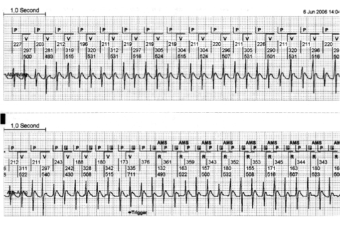
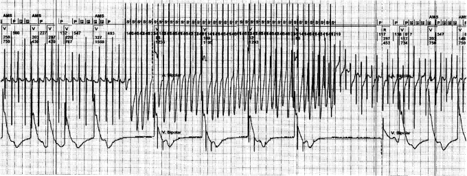
The diagnostic programmes of modern pacemakers have increased our knowledge of atrial tachyarrhythmias (ATAs) in chronically paced patients. These programmes also support the evaluation of the effects of pharmacological treatment of ATAs. The success of interruption and/or prevention of ATAs with pacemakers depends strongly on the diagnostic accuracy and the properties of the pacing algorithms, their individual programming and the site and configuration of the pacing leads. Atrial septum pacing can be beneficial in patients with paroxysmal atrial fibrillation and prolonged P wave duration. Recent large-scale studies on preventive and interruptive atrial pacing of ATAs show modestly positive or no results. Therefore, atrial pacing therapy for ATAs should be considered cautiously, serving as an adjuvant to pharmacological treatment rather than as a primary intervention. This also applies for pacing interventions for ATAs in cardiac resynchronisation therapy. The pacemaker algorithms for the detection of ATAs and atrial lead configuration are crucial for the success of pacemaker-mediated prevention or interruption of ATAs. The success of these interventions is dependant on future improvements of pacemaker technology. (Neth Heart J 2008;16(Suppl1):S20-S24.).

Keywords: atrial fibrillation; atrial pacing; atrial tachycardia; pace intervention; pace prevention.


Figures

Figure 1
Figure 1
Regular atrial tachycardia with a 2:1 lock in. This is a continuous strip from the pacemaker memory. The upper canal of each strip shows the interpretation of the pacemaker of the atrial and ventricular rhythm. The lower canal shows the intracardiac atrial rhythm, derived from the atrial electrode. It is clearly visible that in the beginning of this regular atrial tachycardia, every second atrial depolarisation remains undetected by the pacemaker. This is caused by the programmed postventricular atrial blanking (PVAB). This PVAB (100 msec) was programmed for the prevention of far field R-wave sensing. A slight irregularity of the atrial rhythm in the beginning of the second strip, makes the atrial tachycardia out of the PVAB period and allows the pacemaker to detect all atrial activity. This allows the pacemaker to engage a mode switch to DDI. It is only at this moment that the regular atrial tachycardia can be detected and the window for the implementation of pace interruption is opened. P=sensed atrial activity, P in black box=sensed atrial activity in the refractory period, V=paced ventricular event, R=sensed atrial event. AMS=automatic mode switch.
Figure 2
Figure 2
Termination of rapid atrial flutter with overdrive. The tracing is taken during pacemaker follow-up and shows from top to bottom: (1) top channel surface ECG; (2) the marker channel of the pacemaker (AMS=automated mode switch, A=atrial paced, P=atrial sensed event, P in black box=sensed atrial activity in the ventricular refractory period, V=ventricular paced, R=ventricular sensed activity), s=stimulus of the atrial overdrive; (3) the atrial electrogram sensed between the ring and tip of the atrial lead. The figure depicts an example of a successful manually programmed, fast overdrive pacing by an implanted pacemaker in a patient with a regular atrial tachycardia. The burst shows a 1:1 atrial capture overdriving the regular atrial tachycardia. After the burst, the first beat is a spontaneous atrial activity with intrinsic ventricular activity followed by two paced atrial ventricular sequential beats.
Figure 3
Figure 3
Overdrive pacing modifying atrial tachycardia into atrial fibrillation. The tracing shows from top to bottom: (1) the marker channel of the pacemaker (AMS=automatic mode switch, A=atrial paced, P=atrial sensed event, Pin black box=sensed atrial activity in the refractory period, V=ventricular paced, R=ventricular sensed activity), s=stimulus of the atrial overdrive; (2) the atrial electrogram sensed between the ring and tip of the atrial lead; (3) the ventricular electrogram sensed between the ring and tip of the ventricular lead. The tracing depicts an example of an unsuccessful manually programmed, fast overdrive pacing by an implanted pacemaker in a patient with a regular atrial tachycardia. After the burst of the fast overdrive pacing, atrial fibrillation emerges and mode switch (AMS) to DDIpacing arises. In this case AF is introduced by fast overdrive pacing, stressing its potential risk for initiation ofundesired arrhythmias. Note the irregularity of the first paced ventricular beats prior to onset of overdrive pacing. This pattern is caused by the DDI mode where the pacemaker is sensing some of the atrial activity outside the blanking period, thus causing the irregular ventricular response.

References

    1. Israel CW, Barold SS. Pacemaker systems as implantable cardiac rhythm monitors. Am J Cardio l2001;88:442-5. - PubMed
    1. de Voogt WG, van Hemel NM, van de Bos AA, Koistinen J, Fast JH. Verification of pacemaker automatic mode switching for the detection of atrial fibrillation and atrial tachycardia with Holter recording. Europace 2006;8:950-61. - PubMed
    1. Israel CW, Barold SS. Failure of atrial flutter detection by a pacemaker with a dedicated atrial flutter detection algorithm. Pacing Clin Electrophysiol 2002;25:1274-7. - PubMed
    1. Goethals M, Timmermans W, Geelen P, Backers J, Brugada P. Mode switching failure during atrial flutter: the ‘2:1 lock-in’ phenomenon. Europace 2003;5:95-102. - PubMed
    1. de Voogt W, van Hemel N, Willems A, Visser J, Chitre Y, Bornzin G, et al. Far-field R-wave reduction with a novel lead design: experimental and human results. Pacing Clin Electrophysiol 2005;28:782-8. - PubMed

LinkOut - more resources