. 2009 Feb;26(2):469-79.
doi: 10.1007/s11095-008-9752-7. Epub 2008 Oct 29.

Major SNP (Q141K) variant of human ABC transporter ABCG2 undergoes lysosomal and proteasomal degradations

Affiliations

Major SNP (Q141K) variant of human ABC transporter ABCG2 undergoes lysosomal and proteasomal degradations

Tomoka Furukawa et al. Pharm Res. 2009 Feb.

Abstract

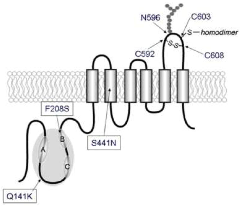
Purpose: Single nucleotide polymorphisms (SNPs) of the ATP-binding cassette (ABC) transporter ABCG2 gene have been suggested to be a significant factor in patients' responses to medication and/or the risk of diseases. We aimed to evaluate the impact of the major non-synonymous SNP Q141K on lysosomal and proteasomal degradations.

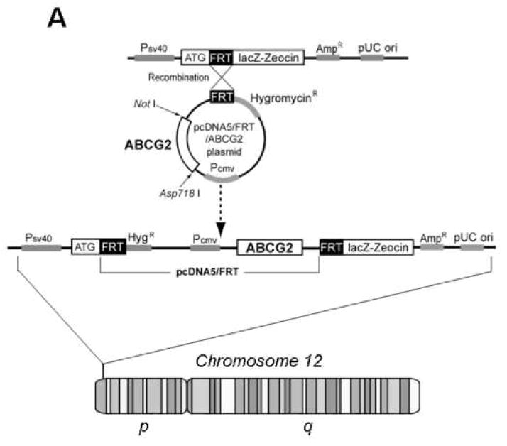
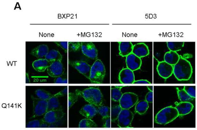
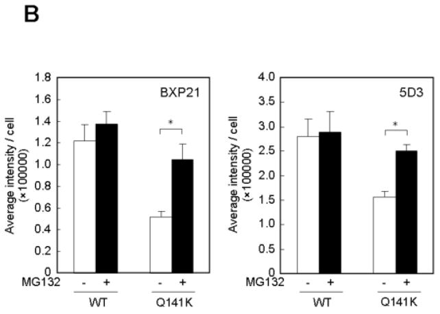
Methods: ABCG2 WT and the Q141K variant were expressed in Flp-In-293 cells by using the Flp recombinase system. Their expression levels and cellular localization was measured by immunoblotting and immunofluorescence microscopy, respectively.

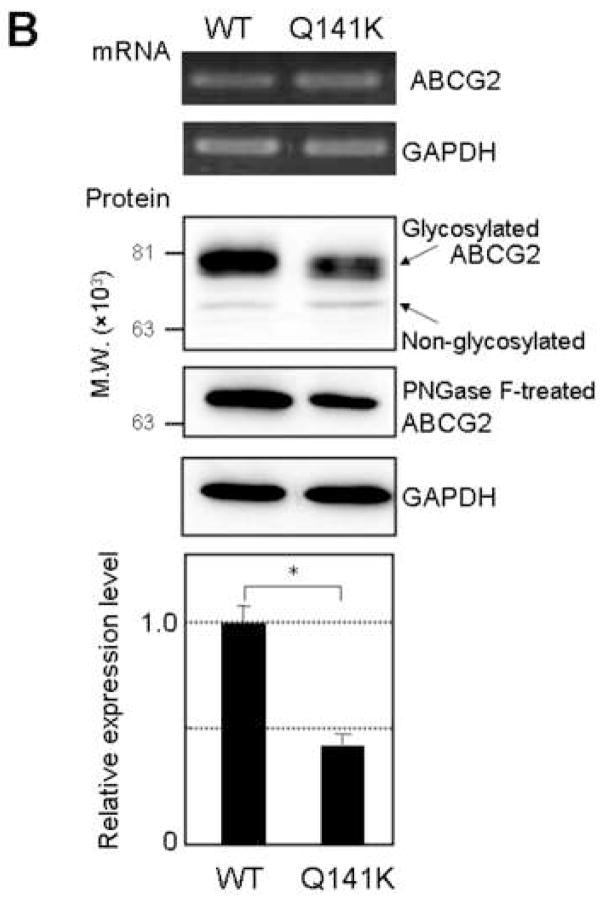
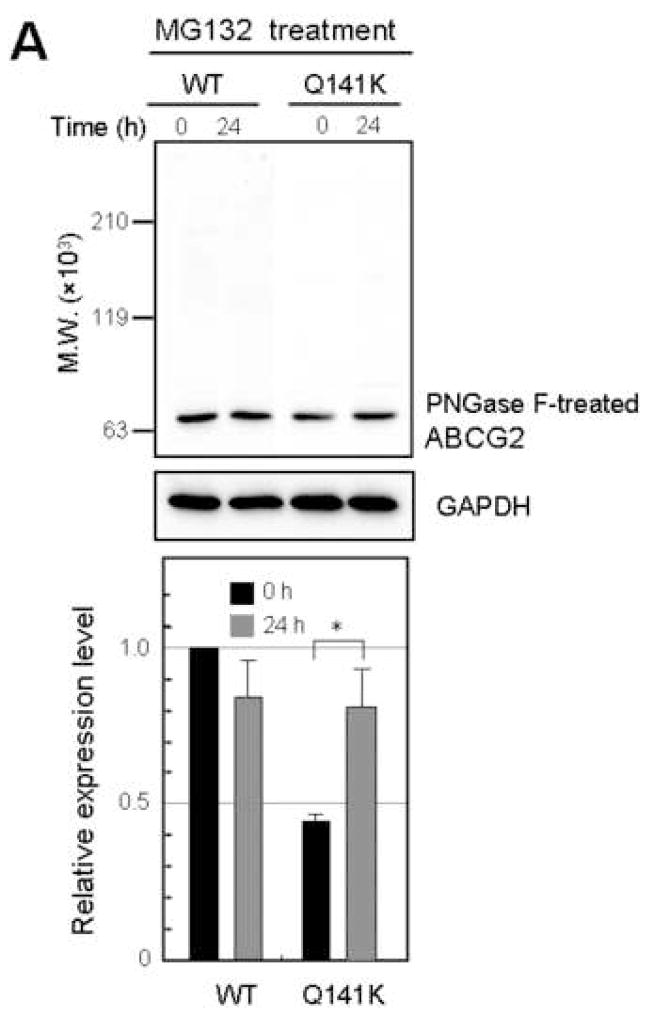
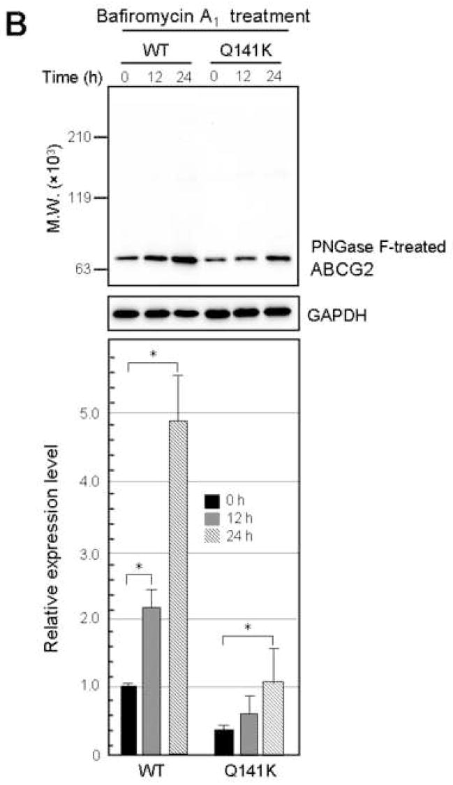
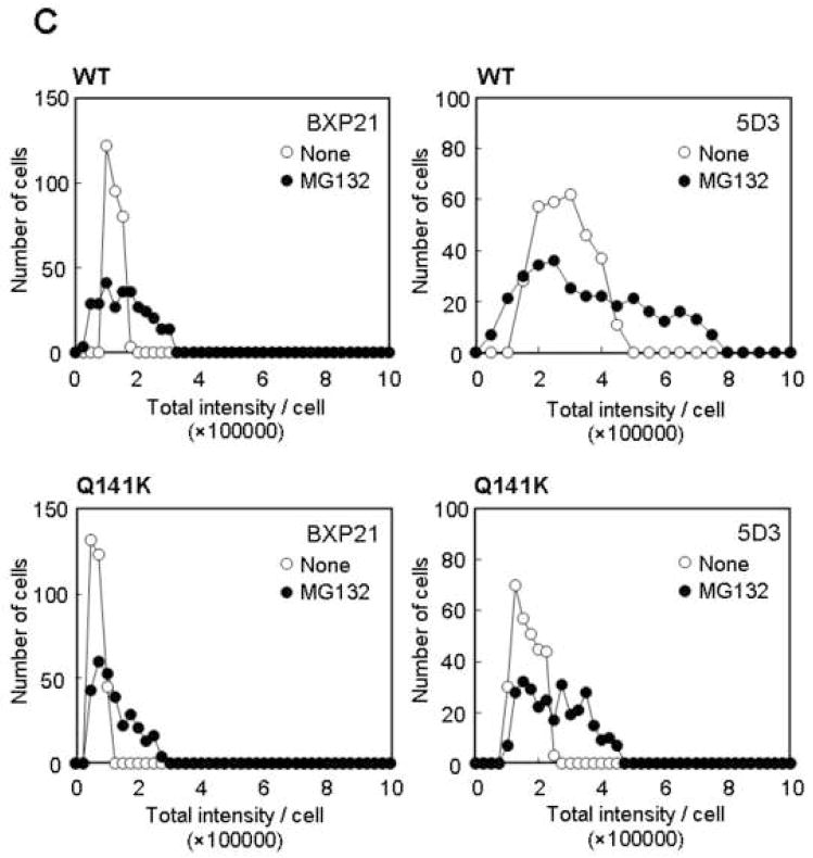
Results: The protein level of the Q141K variant expressed in Flp-In-293 cells was about half that of ABCG2 WT, while their mRNA levels were equal. The protein expression level of the Q141K variant increased about two-fold when Flp-In-293 cells were treated with MG132. In contrast, the protein level of ABCG2 WT was little affected by the same treatment. After treatment with bafilomycin A1, the protein levels of ABCG2 WT and Q141K increased 5- and 2-fold in Flp-In-293 cells, respectively.

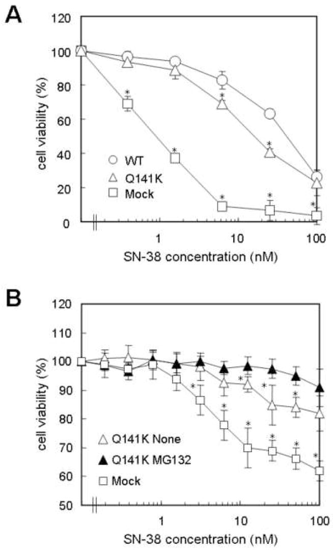
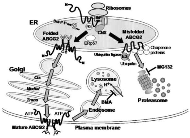
Conclusions: The results strongly suggest that the major non-synonymous SNP Q141K affects the stability of the ABCG2 protein in the endoplasmic reticulum and enhances its susceptibility to ubiquitin-mediated proteasomal degradation.


Figures

Figure 1
Figure 1
Schematic illustration of the protein structure of human ABCG2. The sites of three non-synonymous SNPs, Q141K, F208S and S441N, are indicated. N-linked glycosylation occurs on Asn596 (N596). Cys603 is required for homodimer formation, whereas Cys592 and Cys608 are required for the formation of an intramolecular disulfide bond. A, B, and C indicate the motifs of Walker A (amino acids #80 - 86), Walker B (amino acids #205 - 210), and signature C (amino acids #186 - 200).
Figure 2
Figure 2
Flp-mediated integration of the ABCG2 cDNA into FRT-tagged genomic DNA (A), the mRNA and protein levels of ABCG2 WT and Q141K (B) and the relationship between the relative intensity of ABCG2 immunoreactivity and the amount of cellular proteins (C). A: Genomic DNA of Flp-In-293 cells contains a single integrated Flp recombination target (FRT) site. The cells were co-transfected with the pcDNA5/FRT vector carrying the ABCG2 cDNA and the Flp recombinase expression plasmid pOG44. Flp recombinase mediates insertion of the expression construct with the ABCG2 cDNA into the genome at the integrated FRT site through site-specific DNA recombination. B: The mRNA level was analyzed by RT-PCR with total RNA extracted from Flp-In-293 cells expressing ABCG2 WT or Q141K. For comparison of the protein levels, the cell lysate of each cell population was treated with PNGase F and then analyzed by immunoblotting with the ABCG2-specific monoclonal antibody (BXP-21) or the GAPDH-specific antibody. The level of the Q141K variant protein was normalized to the WT level. C: Relationship between the ABCG2 protein level and the intensity of chemiluminescence. Cell lysate samples (2.5, 5, 10 and 20 μg protein) of Flp-In-293/ABCG2 WT cells were treated with PNGase F and then analyzed by immunoblotting. The signal intensity of ABCG2 was plotted as a function of the logarithmic value of protein.
Figure 2
Figure 2
Flp-mediated integration of the ABCG2 cDNA into FRT-tagged genomic DNA (A), the mRNA and protein levels of ABCG2 WT and Q141K (B) and the relationship between the relative intensity of ABCG2 immunoreactivity and the amount of cellular proteins (C). A: Genomic DNA of Flp-In-293 cells contains a single integrated Flp recombination target (FRT) site. The cells were co-transfected with the pcDNA5/FRT vector carrying the ABCG2 cDNA and the Flp recombinase expression plasmid pOG44. Flp recombinase mediates insertion of the expression construct with the ABCG2 cDNA into the genome at the integrated FRT site through site-specific DNA recombination. B: The mRNA level was analyzed by RT-PCR with total RNA extracted from Flp-In-293 cells expressing ABCG2 WT or Q141K. For comparison of the protein levels, the cell lysate of each cell population was treated with PNGase F and then analyzed by immunoblotting with the ABCG2-specific monoclonal antibody (BXP-21) or the GAPDH-specific antibody. The level of the Q141K variant protein was normalized to the WT level. C: Relationship between the ABCG2 protein level and the intensity of chemiluminescence. Cell lysate samples (2.5, 5, 10 and 20 μg protein) of Flp-In-293/ABCG2 WT cells were treated with PNGase F and then analyzed by immunoblotting. The signal intensity of ABCG2 was plotted as a function of the logarithmic value of protein.
Figure 3
Figure 3
Effect of MG132 (A) or bafilomycin A1 (B) on the protein levels of ABCG2 WT and Q141K variant. A: Flp-In-293 cells expressing ABCG2 WT or Q141K were incubated with 2 μM MG132 for 0 and 24 h. ABCG2 protein levels were analyzed by immunoblotting after PNGase F treatment. B: Flp-In-293 cells expressing ABCG2 WT or Q141K were incubated with 10 nM bafilomycin A1 for 0, 12, and 24 h. ABCG2 protein levels were analyzed as described above. Data are expressed as mean values ± S.D. in triplicate experiments. Statistical significance (*P<0.01) was evaluated by Student’s t-test.
Figure 3
Figure 3
Effect of MG132 (A) or bafilomycin A1 (B) on the protein levels of ABCG2 WT and Q141K variant. A: Flp-In-293 cells expressing ABCG2 WT or Q141K were incubated with 2 μM MG132 for 0 and 24 h. ABCG2 protein levels were analyzed by immunoblotting after PNGase F treatment. B: Flp-In-293 cells expressing ABCG2 WT or Q141K were incubated with 10 nM bafilomycin A1 for 0, 12, and 24 h. ABCG2 protein levels were analyzed as described above. Data are expressed as mean values ± S.D. in triplicate experiments. Statistical significance (*P<0.01) was evaluated by Student’s t-test.
Figure 4
Figure 4
Immunocytochemical staining of Flp-In-293 cells expressing ABCG2 WT or Q141K proteins (A) and statistical data to analyze the effects of MG132 on the cellular localization of ABCG2 WT or Q141K proteins (B, C). A: Cells were incubated with or without 2 μM MG132 for 24 h. ABCG2 proteins were immunologically detected with an ABCG2-specific monoclonal antibody (either BXP-21 or 5D3) and Alexa Fluor 488 (green). Cellular nuclei were stained by Hoechst 33342 (blue), as described in Materials and Methods. The horizontal white bar corresponds to 20 μm. B: Based on the data shown in Figure 4C, the average intensity per cell has been calculated. Data are expressed as mean values ± S.D. (*P<0.005). C: Based on the immunofluorescence microscopic images, the intensity of ABCG2-positive dots was measured for more than 300 cells for each preparation. The number of cells is plotted as a function of total intensity per cell.
Figure 4
Figure 4
Immunocytochemical staining of Flp-In-293 cells expressing ABCG2 WT or Q141K proteins (A) and statistical data to analyze the effects of MG132 on the cellular localization of ABCG2 WT or Q141K proteins (B, C). A: Cells were incubated with or without 2 μM MG132 for 24 h. ABCG2 proteins were immunologically detected with an ABCG2-specific monoclonal antibody (either BXP-21 or 5D3) and Alexa Fluor 488 (green). Cellular nuclei were stained by Hoechst 33342 (blue), as described in Materials and Methods. The horizontal white bar corresponds to 20 μm. B: Based on the data shown in Figure 4C, the average intensity per cell has been calculated. Data are expressed as mean values ± S.D. (*P<0.005). C: Based on the immunofluorescence microscopic images, the intensity of ABCG2-positive dots was measured for more than 300 cells for each preparation. The number of cells is plotted as a function of total intensity per cell.
Figure 5
Figure 5
Drug resistance profiles of Flp-In-293 cells expressing ABCG2 WT or Q141K (A) and the effect of MG132 on the SN-38 resistance of Flp-In-293 cells expressing Q141K (B). A: Cells were incubated with SN-38 at different concentrations for 72 h in the absence of MG132. Cell viability was determined by MTT assay. Data are expressed as mean values ± S.D. in multi-replicated experiments (n = 4). *, P<0.01 as compared with Flp-In-293 cells expressing ABCG2 WT. B: Cells were incubated with SN-38 at different concentrations for 24 h in the presence or absence of 2 μM MG132. Cell viability was determined as described above. Data are expressed as mean values ± S.D. in multi-replicated experiments (n = 8). *, P<0.01 as compared with Flp-In-293 cells expressing ABCG2 Q141K treated with MG132.
Figure 6
Figure 6
Schematic illustration of plausible pathways for protein folding and degradation of ABCG2 WT and Q141K variant protein. BiP(Grp78) is one of the abundant ER chaperones and is closely related to cytosolic Hsp70. Because of its location, BiP can immediately interact with the unfolded nascent peptide chain and hence contribute to the translocation of nascent chains into the ER (49). BiP reportedly has an ATPase domain and a peptide-binding domain that coordinate cycles of ATP hydrolysis and ADP exchange, which stimulate the binding and release of the unfolded protein, respectively (50). During de novo synthesis in the ER, cysteine disulfide bonds are formed and oligosaccharides are added to asparagine (N-glycosylation) or serine residues (O-glycosylation) of glycoproteins. The ER has a unique oxidizing potential that supports disulfide bond formation during protein folding (51). In addition to the well-known oxidoreductases, such as PDI and ERp57, many novel oxidoreductases have been identified over the past few years whose functions and substrates are unknown (52, 53). Some of them are likely to be involved in the formation of disulfide bonds at Cys592, Cys603, and Cys608 in the ABCG2 protein. Furthermore, N-linked glycans are added en block to proteins as “core oligosaccharides” (Glc3Man9GlcNAc2). In the ABCG2 protein, Asn596 is an N-glycosylation site (44, 45). Calnexin (CNX) is located near the translocon and can interact with nascent peptide chains of N-glycosylated proteins. N-linked glycans are subjected to extensive modification as glycoproteins mature and move through the ER via the Golgi apparatus to their final destination, for example the plasma membrane. After remaining in the plasma membrane domain for a certain period, ABCG2 WT is degraded by the endosome-lysosome pathway. In contrast, Q141K undergoes both lysosomal proteolysis and ubiquitination-mediated proteasomal degradation. The proteasomal degradation is inhibited by MG132.

References

    1. Kalow E, Meyer U, Tyndale RF. Pharmacogenomics. Marcel Dekker; New York Besel: 2001.
    1. Ishikawa T, Tamura A, Saito H, Wakabayashi K, Nakagawa H. Pharmacogenomics of the human ABC transporter ABCG2: from functional evaluation to drug molecular design. Naturwissenschaften. 2005:1–13. - PubMed
    1. de Jong FA, de Jonge MJ, Verweij J, Mathijssen RH. Role of pharmacogenetics in irinotecan therapy. Cancer Lett. 2006;234:90–106. - PubMed
    1. Yanase K, Tsukahara S, Mitsuhashi J, Sugimoto Y. Functional SNPs of the breast cancer resistance protein-therapeutic effects and inhibitor development. Cancer Lett. 2006;234:73–80. - PubMed
    1. Backstrom G, Taipalensuu J, Melhus H, Brandstrom H, Svensson AC, Artursson P, Kindmark A. Genetic variation in the ATP-binding cassette transporter gene ABCG2 (BCRP) in a Swedish population. Eur J Pharm Sci. 2003;18:359–64. - PubMed

Publication types

MeSH terms