. 2009 Jan;61(1):43-54.
doi: 10.1007/s00251-008-0335-x. Epub 2008 Oct 29.

Influence of the tapasin C terminus on the assembly of MHC class I allotypes

Affiliations

Influence of the tapasin C terminus on the assembly of MHC class I allotypes

Laura C Simone et al. Immunogenetics. 2009 Jan.

Abstract

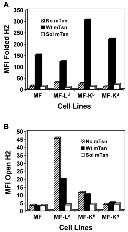
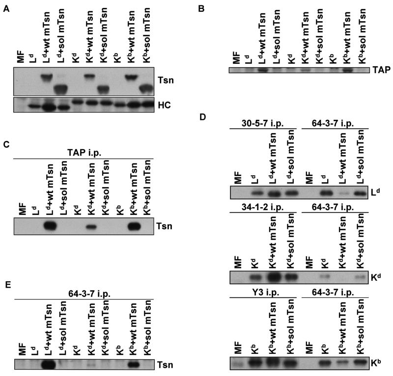
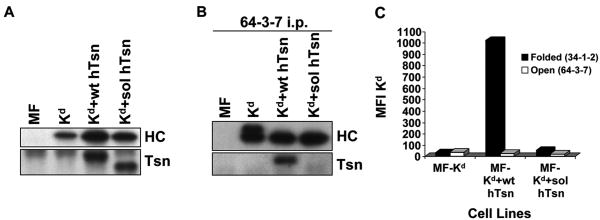
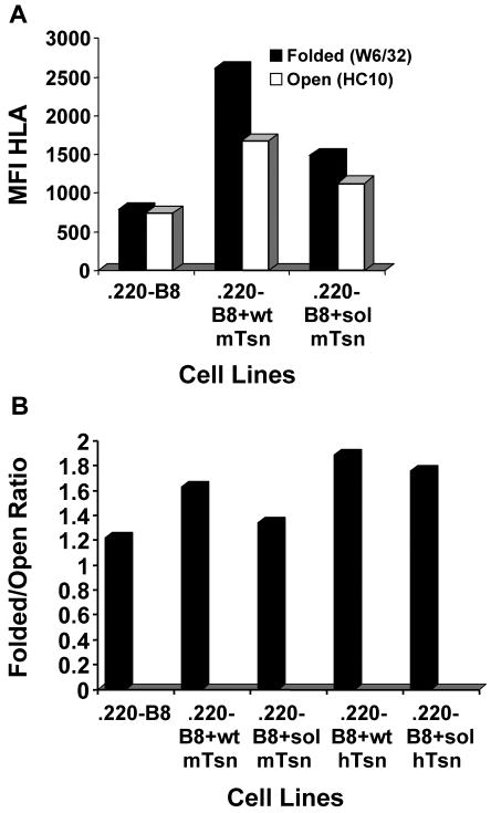
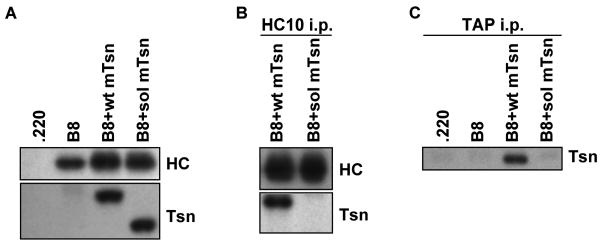
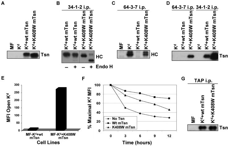
Several endoplasmic reticulum proteins, including tapasin, play an important role in major histocompatibility complex (MHC) class I assembly. In this study, we assessed the influence of the tapasin cytoplasmic tail on three mouse MHC class I allotypes (H2-K(b), -K(d), and -L(d)) and demonstrated that the expression of truncated mouse tapasin in mouse cells resulted in very low K(b), K(d), and L(d) surface expression. The surface expression of K(d) also could not be rescued by human soluble tapasin, suggesting that the surface expression phenotype of the mouse MHC class I molecules in the presence of soluble tapasin was not due to mouse/human differences in tapasin. Notably, soluble mouse tapasin was able to partially rescue HLA-B8 surface expression on human 721.220 cells. Thus, the cytoplasmic tail of tapasin (either mouse or human) has a stronger impact on the surface expression of murine MHC class I molecules on mouse cells than on the expression of HLA-B8 on human cells. A K408W mutation in the mouse tapasin transmembrane/cytoplasmic domain disrupted K(d) folding and release from tapasin, but not interaction with transporter associated with antigen processing (TAP), indicating that the mechanism whereby the tapasin transmembrane/cytoplasmic domain facilitates MHC class I assembly is not limited to TAP stabilization. Our findings indicate that the C terminus of mouse tapasin plays a vital role in enabling murine MHC class I molecules to be expressed at the surface of mouse cells.


Figures

Figure 1
Figure 1
The murine soluble tapasin mutant was not able to facilitate normal H2-Ld, -Kb, and -Kd surface expression. MF, MF+Ld, MF+Kb, or MF+Kd cells transfected with no tapasin, wild type mouse tapasin (Wt mTsn) or soluble mouse tapasin (Sol mTsn) were incubated with secondary antibody only, or with an antibody against the folded form of the transfected mouse MHC class I molecule (30-5-7 for Ld, Y3 for Kb, and 34-1-2 for Kd) or with 64-3-7 against the open form of each epitope-tagged mouse MHC class I molecule. Results obtained with the antibody against the folded form are shown in (A), and results obtained with 64-3-7 are shown in (B). Results obtained with secondary antibody only were all less than 5.0. Values on the y axes are relative mean fluorescence intensity (MFI) units. Very similar results were obtained with independent transfectant clones, with alternative antibodies (B8-24-3 for folded Kb), and in separate flow cytometry experiments.
Figure 2
Figure 2
(A) Western blotting for mouse tapasin showed that soluble tapasin was present at a level similar to (or slightly exceeding) wild type mouse tapasin in transfected MF cells, and Western blotting for the transfected mouse MHC class I heavy chain demonstrated that Ld, Kd, or Kb heavy chains were expressed at a roughly similar level in the MF cells. After electrophoresis of samples of cell lysates on a 10% acrylamide Tris-glycine gel for tapasin or on a 4→20% acrylamide Tris-glycine gel for MHC class I heavy chain, the proteins were blotted and probed with Ab specific for mouse tapasin (Tsn) or with 64-3-7 (for HC). The Ld heavy chain naturally has the 64-3-7 epitope, and the epitope was introduced into the Kd and Kb heavy chains by site-directed mutagenesis. (B) Murine soluble tapasin did not stabilize mouse TAP. Lysate samples were electrophoresed on a 10% acrylamide Tris-glycine gel before blotting and probing with antibody specific for mouse TAP. (C) Soluble mouse tapasin did not associate with TAP. Immunoprecipitates of TAP molecules were electrophoresed on a 10% acrylamide Tris-glycine gel. The proteins were then transferred to a blotting membrane and probed with antiserum specific for mouse tapasin (Tsn). (D) The folding of Ld, Kd, and Kb heavy chains was improved by the intracellular presence of wild type tapasin but not soluble tapasin. Immunoprecipitations were performed with mAb 30-5-7, 34-1-2, and Y3 (for folded Ld, Kd, and Kb, respectively) on lysates of MF cells that expressed each of the aforementioned MHC class I heavy chains along with no tapasin, wild type tapasin, or soluble tapasin. Immunoprecipitations with the 64-3-7 mAb were also done on corresponding cell lysates. The immunoprecipitates were electrophoresed on 4→20% acrylamide Tris-glycine gels, transferred to membranes, and probed with 64-3-7. (E) Tapasin association with the mouse MHC class I heavy chain was abrogated by the deletion of the tapasin TM/CYT region. Open MHC class I heavy chains were immunoprecipitated from a lysate of each of the indicated cell types with the 64-3-7 mAb. The immunoprecipitates were electrophoresed on a 10% acrylamide Tris-glycine gel, transferred to a membrane, and probed with an anti-tapasin antibody (Tsn).
Figure 3
Figure 3
(A) Tapasin and Kd were expressed at similar levels in Kd+wt hTsn relative to Kd+sol hTsn. Samples of lysates from MF, MF+Kd, MF+Kd+wt hTsn, and MF+Kd+sol hTsn were electrophoresed on a 10% acrylamide Tris-glycine gel for tapasin (Tsn) or on a 4→20% acrylamide Tris-glycine gel for Kd, the proteins were transferred to a blotting membrane and probed with Ab specific for human tapasin (Tsn) or with 64-3-7 to identify the total epitope-tagged Kd heavy chain (HC). (B) Soluble human tapasin binds very weakly to Kd. Open Kd heavy chains were immunoprecipitated from a digitonin lysate of each of the indicated cell types with the 64-3-7 mAb in the presence of 200 μM DSP. The immunoprecipitates were electrophoresed on acrylamide Tris-glycine gels, transferred to membranes, and probed with 64-3-7 to identify the immunoprecipitated open Kd heavy chain (HC) or with anti-tapasin mAb (Tsn) to identify the co-immunoprecipitated tapasin. (C) Soluble human tapasin did not enable Kd to be expressed at the surface of mouse cells. Cells were incubated with secondary antibody only, or an antibody against the folded form of Kd (34-1-2), or an antibody against the open form of Kd (64-3-7). Results obtained with the secondary antibody only were all less than 5.0. Values on the y axis are relative mean fluorescence intensity (MFI) units. Wt hTsn = wild type human tapasin; Sol hTsn = soluble human tapasin. Very similar results were obtained in a separate flow cytometry experiment.
Figure 4
Figure 4
Soluble mouse tapasin partially restored B8 expression at the surface of 721.220 cells. (A) Cells (.220-B8, .220-B8+wt mTsn, and .220-B8+sol mTsn) were incubated with secondary antibody only (data not shown), or with an antibody against the folded form of the HLA class I molecule (W6/32), or with an antibody against the open form of the HLA class I molecule (HC10). Results obtained with the secondary antibody only were all less than 11.0. Values on the y axis are relative mean fluorescence intensity (MFI) units. Wt mTsn = wild type mouse tapasin; Sol mTsn = soluble mouse tapasin. Very similar results were obtained in a separate flow cytometry experiment. (B) Cells (.220-B8, .220-B8+wt mTsn, .220-B8+sol mTsn, .220-B8+wt hTsn, .220-B8+sol hTsn) were incubated with secondary antibody only, W6/32, or HC10. Results obtained with the secondary antibody only were all less than 7.0. The ratio of the mean fluorescence intensity for W6/32 versus HC10 for each cell type is displayed.
Figure 5
Figure 5
(A) Tapasin and B8 were expressed at similar levels in B8+wt mTsn relative to B8+sol mTsn. Samples of lysates from .220, .220-B8, .220-B8+wt mTsn, and .220-B8+sol mTsn were electrophoresed on a 10% acrylamide Tris-glycine gel for tapasin (Tsn) or on a 4→20% acrylamide Tris-glycine gel for B8, the proteins were transferred to a blotting membrane and probed with Ab specific for mouse tapasin (Tsn) or with HC10 to identify the open B8 heavy chain (HC). (B) Soluble mouse tapasin does not associate with B8. Open B8 heavy chains were immunoprecipitated from a lysate of each of the indicated cell types with the HC10 mAb. The immunoprecipitates were electrophoresed on acrylamide Tris-glycine gels, transferred to membranes, and probed with HC10 to identify the immunoprecipitated open B8 heavy chain (HC) or with anti-mouse tapasin antibody (Tsn) to identify the co-immunoprecipitated tapasin. (C) Soluble mouse tapasin does not associate with human TAP. TAP1 was immunoprecipitated from cell lysates with antibody 148.3. After electrophoresis of the immunoprecipitated proteins on a 10% acrylamide Tris-glycine gel, the proteins were blotted and probed with anti-mouse tapasin antibody (Tsn).
Figure 6
Figure 6
(A) Tapasin K408W was detected in similar or slightly greater amount than wild type tapasin in the transfected cells. Samples of cell lysates were electrophoresed on a 10% acrylamide Tris-glycine gel, and the proteins were transferred to a blotting membrane and probed with an antibody for mouse tapasin (Tsn). (B) A majority of the Kd molecules folded in the presence of mouse tapasin K408W were Endo H sensitive. Folded Kd molecules were immunoprecipitated with 34-1-2, electrophoresed on 4→20% acrylamide Tris-glycine gel, blotted, and probed with 64-3-7 (HC). Samples were treated (+) or untreated (-) with Endo H as indicated. (C) Mouse tapasin K408W was found to increase the amount of Kd in the open conformation. Immunoprecipitations were performed with antibody 64-3-7 on lysates of the indicated cell types. The immunoprecipitated proteins were electrophoresed on a 4→20% acrylamide Tris-glycine gel, transferred, and probed on a blot with 64-3-7 (HC). (D) Kd was more strongly associated with tapasin K408W than with wild type tapasin, and both wild type tapasin and tapasin K408W were predominantly associated with the folded, and not the open, form of Kd. Immunoprecipitations were performed with mAb 64-3-7 and 34-1-2 on lysates of the indicated cell types. The immunoprecipitated proteins were electrophoresed on a 10% acrylamide Tris-glycine gel, transferred to a membrane, and probed with anti-tapasin antibody (Tsn). (E) The amount of open Kd at the cell surface was increased by expression of tapasin K408W compared to wild type tapasin. Cells were incubated with secondary antibody only, or with an antibody against the open form of the Kd molecule (64-3-7). Background staining with PE-conjugated secondary antibody only was <5.0 for both MF-Kd+wt mTsn and MF-Kd+K408W mTsn (not shown). Values on the y axis are relative mean fluorescence intensity (MFI) units. Similar results were obtained in a separate flow cytometry experiment. (F) Kd molecules assembled in cells expressing mouse tapasin K408W were less stable than those assembled in the presence of wild type mouse tapasin. Cells were treated with 2 μg/ml Bfa in complete medium for 0, 3, 6, 9, or 12 hours, then stained with 34-1-2 and analyzed by flow cytometry. (G) The K408W mutation in mouse tapasin does not abrogate the association of tapasin with TAP. Immunoprecipitations were performed with an anti-TAP antiserum on lysates of the cell lines indicated. The immunoprecipitated proteins were electrophoresed on a 10% acrylamide Tris-glycine gel, and probed on a Western blot with an anti-mouse tapasin antibody.

Similar articles

Cited by

References

    1. Allen H, Wraith D, Pala P, Askonas B, Flavell RA. Domain interactions of H-2 class I antigens alter cytotoxic T-cell recognition sites. Nature. 1984;309:279–281. - PubMed
    1. Bangia N, Lehner PJ, Hughes EA, Surman M, Cresswell P. The N-terminal region of tapasin is required to stabilize the MHC class I loading complex. Eur J Immunol. 1999;29:1858–1870. - PubMed
    1. Barnden MJ, Purcell AW, Gorman JJ, McCluskey J. Tapasin-mediated retention and optimization of peptide ligands during the assembly of class I molecules. J Immunol. 2000;165:322–330. - PubMed
    1. Carreno BM, Hansen TH. Exogenous peptide ligand influences the expression and half-life of free HLA class I heavy chains ubiquitously detected at the cell surface. Eur J Immunol. 1994;24:1285–1292. - PubMed
    1. Carreno B, Solheim JC, Harris M, Stroynowski I, Connolly JM, Hansen TH. TAP associates with a unique class I conformation, whereas calnexin associates with multiple class I forms in mouse and man. J Immunol. 1995;155:4726–4733. - PubMed

Publication types

MeSH terms