. 2009 Mar;4(1):129-39.
doi: 10.1007/s11481-008-9128-0. Epub 2008 Oct 29.

Methamphetamine enhances HIV-1 infectivity in monocyte derived dendritic cells

Affiliations

Methamphetamine enhances HIV-1 infectivity in monocyte derived dendritic cells

Madhavan P N Nair et al. J Neuroimmune Pharmacol. 2009 Mar.

Abstract

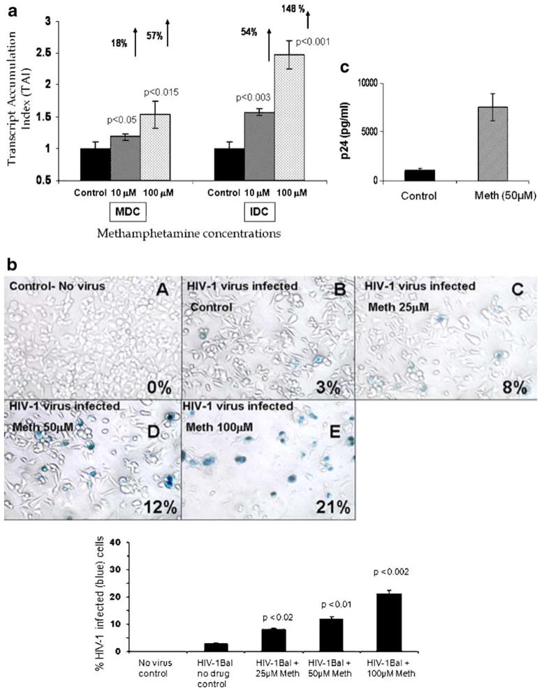
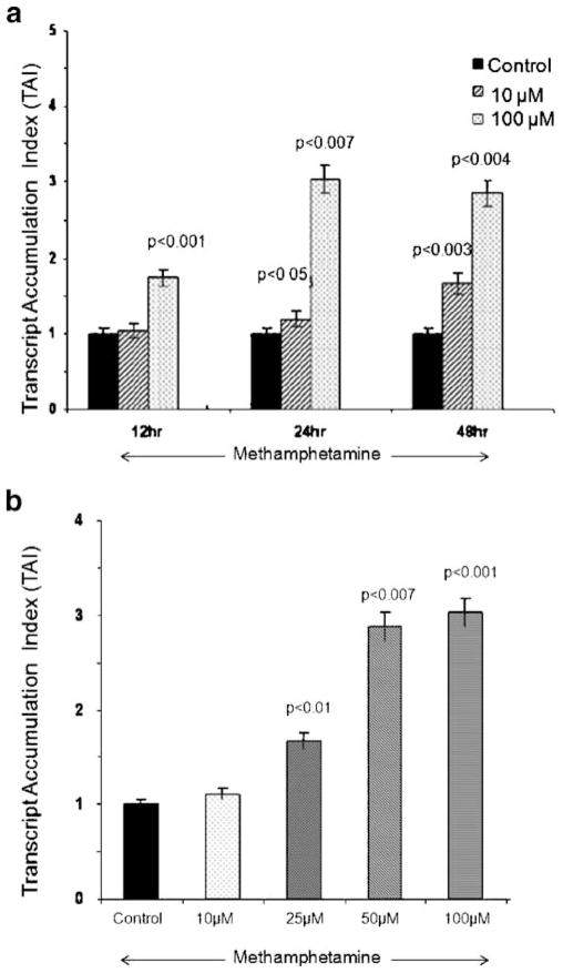
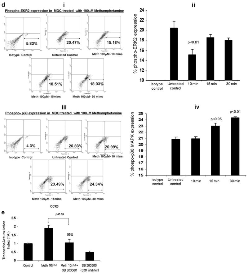
The US is currently experiencing an epidemic of methamphetamine (Meth) use as a recreational drug. Recent studies also show a high prevalence of HIV-1 infection among Meth users. We report that Meth enhances HIV-1 infectivity of dendritic cells as measured by multinuclear activation of a galactosidase indicator (MAGI) cell assay, p24 assay, and LTR-RU5 amplification. Meth induces increased HIV-1 infection in association with an increase in the HIV-1 coreceptors, CXCR4 and CCR5, and infection is mediated by downregulation of extracellular-regulated kinase (ERK2) and the upregulation of p38 mitogen-activated protein kinase (MAPK). A p38 inhibitor (SB203580) specifically reversed the Meth-induced upregulation of the CCR5 HIV-1 coreceptor. The dopamine D2 receptor antagonist RS +/- sulpiride significantly reversed the Meth-induced upregulation of CCR5, demonstrating that the Meth-induced effect is mediated via the D2 receptor. These studies report for the first time that Meth fosters HIV-1 infection, potentially via upregulating coreceptor gene expression. Further, Meth mediates its regulatory effects via dopamine receptors and via downregulating ERK2 with a reciprocal upregulation of p38 MAPK. Elucidation of the role of Meth in HIV-1 disease susceptibility and the mechanism through which Meth mediates its effects on HIV-1 infection may help to devise novel therapeutic strategies against HIV-1 infection in high-risk Meth-using HIV-1-infected subjects.


Figures

Fig. 1
Fig. 1
a Meth enhances HIV-1 replication. MDC and IDC (5×10⁵ cells per milliliter) were infected with native HIV-1 IIIB [X4] (NIH AIDS Research and Reference Reagent Program Cat# 398) at a concentration of 103.0 TCID50 per milliliter cells overnight and washed three times with Hank’s balanced salt solution (GIBCO-BRL, Grand Island, NY, USA) before being returned to culture with and without Meth (10 and 100 μM) for 24 h. The RNA was extracted, reverse-transcribed, and followed by quantitative real-time PCR against the LTR-RU5 and the housekeeping gene, β-actin, and the 18S RNA primers as internal controls. The data represent mean±SD of three independent experiments. Statistical analysis was done using Student’s t test. b Meth enhances HIV-1 infectivity as measured by MAGI assay. The MAGI cells are HeLa-derived cells stably transfected with CD4 and a reporter construct consisting of the β-galactosidase gene (which is modified to localize to the nucleus) driven by a truncated HIV-1 LTR. Expression of the β-galactosidase gene is Tat dependent such that an incoming virus must produce active Tat protein to drive expression of the reporter. HIV-1-infected cells stained blue with the X-Gal dye. MAGI cells (4×10⁴ cells per well) were plated in a 24-well plate and were treated in duplicate with a 100-μl supernatants from MDCs that have been infected with HIV-1 IIIB virus and treated with or without Meth. A total of 200 μl of DMEM supplemented with 10% FCS, 100 U/ml penicillin, 100 μg/ml streptomycin, 0.25 μg/ml Fungizone, and 300 μg/ml glutamine containing DEAE-DEXTRAN at a concentration of 15 mg/ml was added to the MAGI cells and infected cells were incubated for 3 days at 37°C, 5%CO2. Cells were fixed and stained with 5-bromo-4-chloro-3-indolyl-D-galactopyranoside (X-Gal) and blue cells were counted as infected cells. The graph represents the percentage of infected cells in each treatment group as observed using a ×20 inverted Nikon microscope. c Meth enhances p24 production. MDCs (5×10⁵ cells per milliliter) from normal subjects were infected with native HIV-1 IIIB (NIH AIDS Research and Reference Reagent Program Cat# 398) at a concentration of 103.0 TCID50 per milliliter cells overnight and washed three times with Hank’s balanced salt solution (GIBCO-BRL, Grand Island, NY, USA) before being returned to culture with and without Meth (50 μM) for 14 days. The culture supernatants were quantitated for p24 antigen using a p24 ELISA kit (ZeptoMetrix Corporation, Buffalo, NY, USA). The data represent the mean±SD of three independent experiments and are expressed as picogram per milliliter. Statistical analysis was done using Student’s t test
Fig. 2
Fig. 2
a Meth upregulates CXCR4 gene expression. MDCs (5×10⁵ cells per milliliter) were cultured with Meth (10–100 μM) for 12–48 h; RNA was extracted, reverse-transcribed, and PCR-amplified against a CXCR4 primer using quantitative real-time PCR. The data represent mean±SD of three independent experiments. Statistical analysis was done using Student’s t test. b Meth upregulates CCR5 gene expression. MDCs (5×10⁵ cells per milliliter) were cultured with Meth (10–100 μM) for 24 h; RNA was extracted, reverse-transcribed, and PCR-amplified against a CCR5 primer using quantitative real-time PCR. The data represent mean±SD of three independent experiments. Statistical analysis was done using Student’s t test
Fig. 3
Fig. 3
a Meth differentially regulates signal transduction molecules in MDC. MDCs (5×10⁵ cells per milliliter) were cultured for 24 h with and without Meth (10 and 100 μM); RNA was extracted, reverse-transcribed, and QPCR-amplified against various signal transduction molecules ERK2 and p38 MAPK. The data represent mean±SD of three independent experiments. Statistical significance was determined by Student’s t test. b Meth suppresses ERK2 protein expression in MDC. MDCs were cultured with Meth (10 and 100 μM) for 72 h; proteins were extracted and ERK2 expression was detected by Western blot analysis using an ERK2-specific antibody (Santacruz Biotech Inc). ERK2 migrates as a 44-kDa band on a 4–20% SDS-PAGE. The graphical representation is of the densitometric quantitation of the protein signal expressed as percent change in OD with respect to the untreated control (n=2). Statistical significance was determined by Student’s t test. c Meth increased p38 MAPK protein expression in MDC. MDCs were cultured with Meth (100 μM) for 72 h; proteins were extracted and p38 expression was detected by Western blot analysis using a p38 MAPK-specific antibody (Santacruz Biotech Inc). p38 migrates as a 40-kDa band on a 4–20% SDS-PAGE. The graphical representation is of the densitometric quantitation of the protein signal expressed as percent change in OD with respect to the untreated control (n=2). Statistical significance was determined by Student’s t test. d (i–iv) Effect of Meth on the phosphorylation of p38 MAPK and ERK2. MDCs (~5×10⁵ cell per milliliter) were treated with Meth (100 μM) for 10, 15, and 30 min following FACS analysis which was done to determine the percentage of cells expressing phosphorylated ERK2 and p38 MAPK. Statistical significance was calculated by Student’s t test. The FACS plots are from a representative experiment, while the histogram is a graphical representation of three independent FACS experiments. e p38 MAPK inhibitor reverses Meth-induced upregulation of CCR5 gene expression. MDCs (5×10⁵ cells per milliliter) were cultured alone or with Meth (10 μM) or the p38 MAPK inhibitor (SB20358; 10 μM) alone and in combination with Meth (10 μM) plus inhibitor (SB20358; 10 μM) for 24 h and CCR5-specific mRNA expression was quantitated using real-time PCR. The data represent mean±SD of two independent experiments. Statistical analysis was done using ANOVA
Fig. 3
Fig. 3
a Meth differentially regulates signal transduction molecules in MDC. MDCs (5×10⁵ cells per milliliter) were cultured for 24 h with and without Meth (10 and 100 μM); RNA was extracted, reverse-transcribed, and QPCR-amplified against various signal transduction molecules ERK2 and p38 MAPK. The data represent mean±SD of three independent experiments. Statistical significance was determined by Student’s t test. b Meth suppresses ERK2 protein expression in MDC. MDCs were cultured with Meth (10 and 100 μM) for 72 h; proteins were extracted and ERK2 expression was detected by Western blot analysis using an ERK2-specific antibody (Santacruz Biotech Inc). ERK2 migrates as a 44-kDa band on a 4–20% SDS-PAGE. The graphical representation is of the densitometric quantitation of the protein signal expressed as percent change in OD with respect to the untreated control (n=2). Statistical significance was determined by Student’s t test. c Meth increased p38 MAPK protein expression in MDC. MDCs were cultured with Meth (100 μM) for 72 h; proteins were extracted and p38 expression was detected by Western blot analysis using a p38 MAPK-specific antibody (Santacruz Biotech Inc). p38 migrates as a 40-kDa band on a 4–20% SDS-PAGE. The graphical representation is of the densitometric quantitation of the protein signal expressed as percent change in OD with respect to the untreated control (n=2). Statistical significance was determined by Student’s t test. d (i–iv) Effect of Meth on the phosphorylation of p38 MAPK and ERK2. MDCs (~5×10⁵ cell per milliliter) were treated with Meth (100 μM) for 10, 15, and 30 min following FACS analysis which was done to determine the percentage of cells expressing phosphorylated ERK2 and p38 MAPK. Statistical significance was calculated by Student’s t test. The FACS plots are from a representative experiment, while the histogram is a graphical representation of three independent FACS experiments. e p38 MAPK inhibitor reverses Meth-induced upregulation of CCR5 gene expression. MDCs (5×10⁵ cells per milliliter) were cultured alone or with Meth (10 μM) or the p38 MAPK inhibitor (SB20358; 10 μM) alone and in combination with Meth (10 μM) plus inhibitor (SB20358; 10 μM) for 24 h and CCR5-specific mRNA expression was quantitated using real-time PCR. The data represent mean±SD of two independent experiments. Statistical analysis was done using ANOVA
Fig. 4
Fig. 4
a Effects of D1R-specific siRNA (AF498961) on D1 receptor gene expression in siRNA-transfected MDC. siRNA against the D1 receptor was designed using siRNA design software available on the Invitrogen Web site. The D1 receptor gene (accession no. AF498961) siRNA sequences were as follows, AAGUUGGUCACCUUGGACC[dt][dt], and transfection was done using Lipofectamine reagent. Our results show that D1R siRNA transfection (60 pM) was transient and silencing of D1R gene was evident at 24 h (85% inhibition of D1R gene expression) posttransfection. Data represent mean±SD of three separate experiments and statistical analysis was done using Student’s t test (n=3). b Effects of D1R-specific siRNA on CCR5 gene expression in siRNA-transfected MDC. MDCs were transfected with siRNA using Lipofectamine reagent. Our results show that Meth-treated D1R siRNA-transfected cells showed a significant reduction in CCR5 gene expression indicating that the effect of Meth may be mediated via the D1 receptor. Data represent mean±SD of two separate experiments and statistical analysis was done using ANOVA. c D2 receptor antagonist reverses Meth-induced upregulation of CCR5 gene expression. MDCs were cultured with and without the D2 receptor antagonist RS ± sulpiride (100 μM) alone and in combination with Meth (10 μM) for 24 h and the CCR5-specific mRNA expression was quantitated using real-time PCR. The data represent mean±SD of three independent experiments. Statistical analysis was done using Student’s t test

References

    1. Baselt RC. Disposition of toxic drugs and chemicals in man. 2. Vol. 38. Biomedical Publications; Davis: 1982. p. 113.
    1. Blauvelt A, Asada HM, Saville W, Klaus-Kovtun V, Altman DJ, Yarchoan R. Productive infection of dendritic cells by HIV-1 and their ability to capture virus are mediated through separate pathways. J Clin Invest. 1997;100:2043–2053. doi: 10.1172/JCI119737. - DOI - PMC - PubMed
    1. Cameron P, Pope M, Granelli-Piperno A, Steinman RM. Dendritic cells and the replication of HIV-1. J Leukoc Biol. 1996;59:158–171. - PubMed
    1. Cao H, Verge V, Baron C, Martinache C, Leon A, Scholl S. In vitro generation of dendritic cells from human blood monocytes in experimental conditions compatible for in vivo cell therapy. J Hematother Stem Cell Res. 2000;9:183–194. doi: 10.1089/152581600319397. - DOI - PubMed
    1. Caux C, Massacrier C, Vanbervliet B, Dubois B, Van Kooten C, Durand I. Activation of human DC through CD40 cross-linking. J Exp Med. 1994;180:1263–1272. doi: 10.1084/jem.180.4.1263. - DOI - PMC - PubMed

Publication types

MeSH terms