. 2009 Apr;19(4):321-37.
doi: 10.1002/hipo.20516.

A role for hilar cells in pattern separation in the dentate gyrus: a computational approach

Affiliations

A role for hilar cells in pattern separation in the dentate gyrus: a computational approach

Catherine E Myers et al. Hippocampus. 2009 Apr.

Abstract

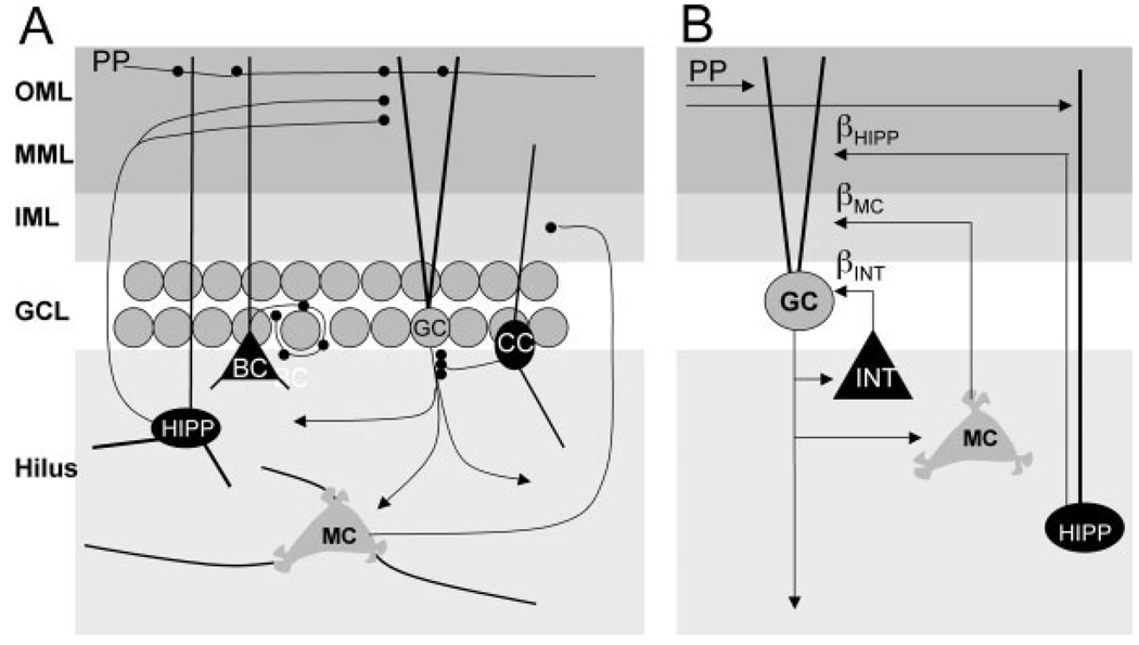
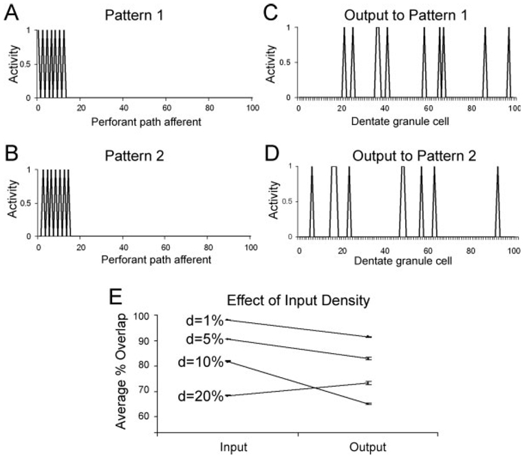
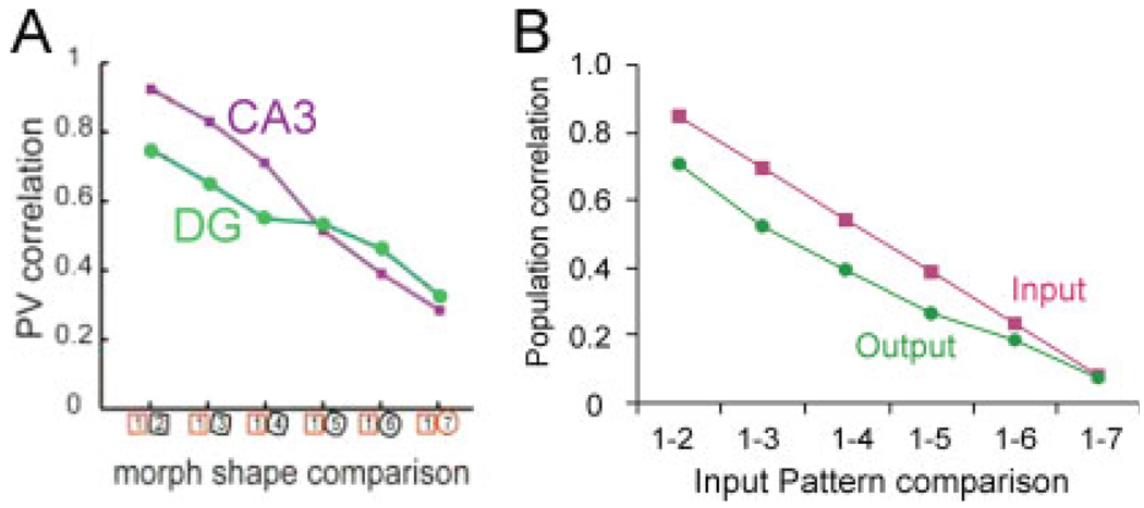
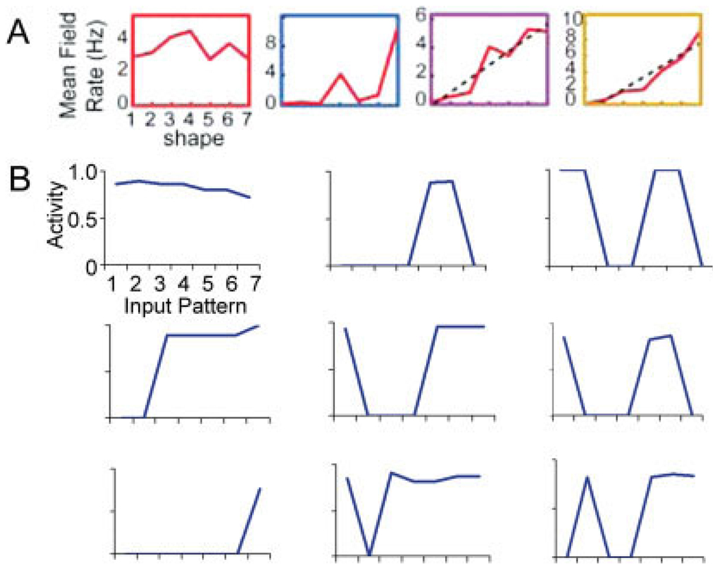
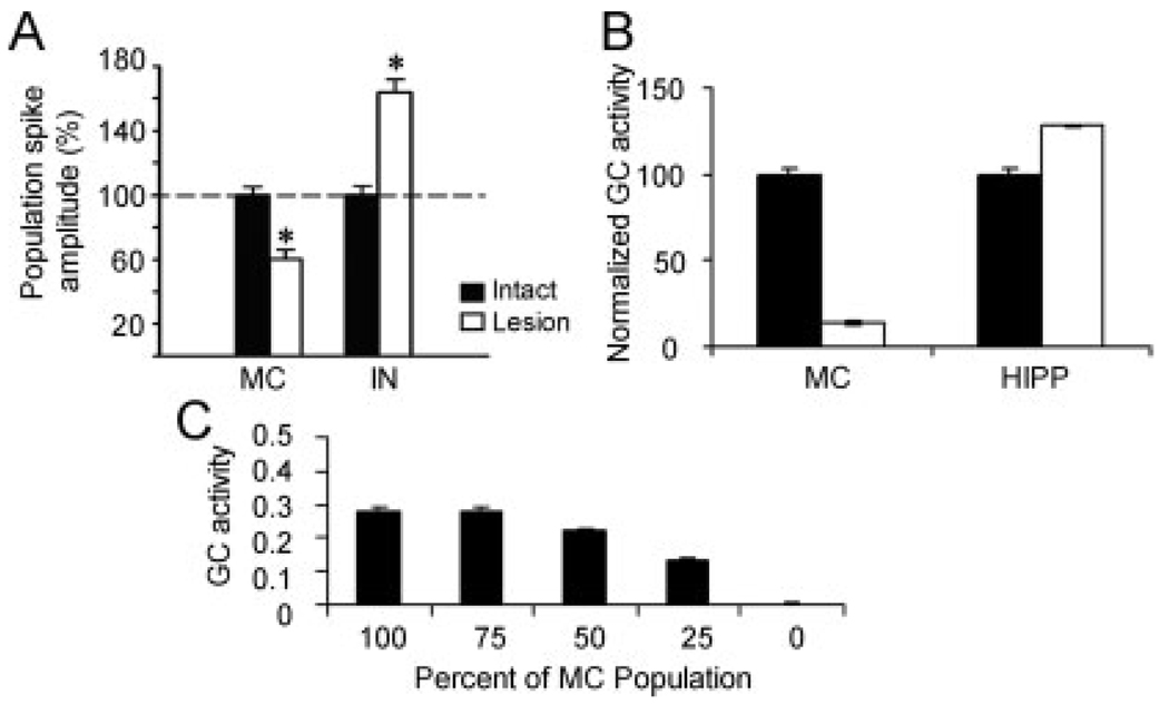
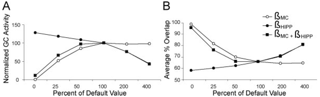
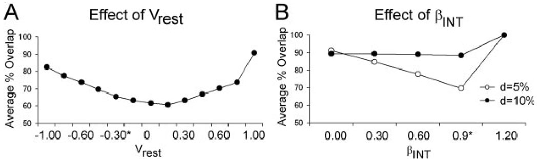
We present a simple computational model of the dentate gyrus to evaluate the hypothesis that pattern separation, defined as the ability to transform a set of similar input patterns into a less-similar set of output patterns, is dynamically regulated by hilar neurons. Prior models of the dentate gyrus have generally fallen into two categories: simplified models that have focused on a single granule cell layer and its ability to perform pattern separation, and large-scale and biophysically realistic models of dentate gyrus, which include hilar cells, but which have not specifically addressed pattern separation. The present model begins to bridge this gap. The model includes two of the major subtypes of hilar cells: excitatory hilar mossy cells and inhibitory hilar interneurons that receive input from and project to the perforant path terminal zone (HIPP cells). In the model, mossy cells and HIPP cells provide a mechanism for dynamic regulation of pattern separation, allowing the system to upregulate and downregulate pattern separation in response to environmental and task demands. Specifically, pattern separation in the model can be strongly decreased by decreasing mossy cell function and/or by increasing HIPP cell function; pattern separation can be increased by the opposite manipulations. We propose that hilar cells may similarly mediate dynamic regulation of pattern separation in the dentate gyrus in vivo, not only because of their connectivity within the dentate gyrus, but also because of their modulation by brainstem inputs and by the axons that "backproject" from area CA3 pyramidal cells.


Figures

FIGURE 1
FIGURE 1
(A) Schematic of some major cell types and connections in the dentate gyrus. Granule cell (GC) bodies lie in the granule cell layer (GCL), with dendrites extending through the inner molecular layer (IML), middle molecular layer (MML), and outer molecular layer (OML), where they receive afferents from the entorhinal cortex layer II neurons via the perforant path (PP). GC axons form the mossy fibers, which synapse in area CA3 (not shown) and also ramify widely within the hilus, synapsing on cell types including the mossy cells (MC), which project primarily to the inner molecular layer, and hilar interneurons. One subtype of hilar interneuron that is prominent in the model is the HIPP cell, which has a hilar cell body and axon that projects primarily to the outer two-thirds of the molecular layer, the location where the perforant path terminates. HIPP cell dendrites extend into the molecular layer, where they are targeted by the PP. Other GABAergic cell types include basket cells (BC), which project to GC bodies, and chandelier cells (CC), which target the axon initial segment of GCs. (B) Elements incorporated in the computational model include GCs, which receive input from the PP and whose axons provide the principal output of the network; interneurons (INT) that innervate GCs; MCs, which are excited by GCs and provide widely ramifying feedback to GCs across the network; and HIPP cells, which receive PP input and inhibit GCs. The global influence of INT, hilar MCs, and HIPP cells on GCs is specified in the model by three constants, βINT, βMC, and βHIPP, which can be modified to simulate upregulation or downregulation of the effect of each cell type on GC activity.
FIGURE 2
FIGURE 2
Example of input and output patterns in the dentate gyrus model. (A) and (B) show two input patterns, presented as activity along the 100 perforant path afferents. Each pattern has seven active perforant path afferents, six of which are common between the two patterns; hence the two patterns are highly overlapping. (C) and (D) show the output, as granule cell activity, to each input pattern in a sample simulation run. For clarity, only the first 100 granule cells are shown; these 100 cells were representative of the larger sample of 500 granule cells in the model. In both (C) and (D), a similar percentage of granule cells become active, but the particular granule cells responding to each pattern differ. Thus, there is less overlap in the outputs than was initially present in the inputs—and so pattern separation has occurred. (E) Results of the model, trained on sets of 10 randomly constructed input patterns, for various densities d of perforant path activation. Pattern separation occurs if the overlap, measured at the granule cell outputs, is less than the overlap measured at the inputs. For low-to-moderate input density (d ≤ 10%), average percent overlap computed at the granule cell outputs is higher than the overlap of the inputs—meaning that pattern separation has occurred. For denser input patterns (d = 20%), the percent overlap measured at the outputs is actually higher than that of the inputs—meaning that pattern separation has failed.
FIGURE 3
FIGURE 3
Simulation of the Leutgeb et al. (2007) “morphed environments.” (A) To simulate the “morphed environments,” a set of overlapping input patterns was constructed. These patterns are then presented, one at a time, as inputs to the dentate gyrus model. (B) Average percent overlap for the input patterns shown in A was highest for “neighboring” patterns such as Patterns 1 and 2, and progressively less for more distinct pairs of patterns such as the extreme Patterns 1 and 7. Average percent overlap, computed across granule cell outputs in the model, was strongly decreased for the most overlapping inputs (e.g., Patterns 1 and 2 or Patterns 1 and 3), but there was less decrease for patterns that were already fairly distinct—and actually a slight increase in overlap for the extremely different input Patterns 1 and 7. Thus, pattern separation in the model is greatest for inputs that overlap extensively.
FIGURE 4
FIGURE 4
(A) In rats, population responses in CA3 are highly correlated in neighboring environments, and this correlation decreases approximately linearly as environmental difference increases; in contrast, there is less correlation in dentate gyrus (DG) responses, particularly for environments that are very similar. Adapted from Leutgeb et al. (2007), Figure 2C. (B) Population responses from dentate granule cells in the model (“Output”) also show reduced correlation for neighboring stimuli, compared with correlations that exist in the input patterns (“Input”). [Color figure can be viewed in the online issue which is available at www.interscience.wiley.com.]
FIGURE 5
FIGURE 5
(A) Mean responses from representative dentate granule cells in rats as the environment is progressively “morphed” through seven stages; response curves can be linear, sigmoidal, and even biphasic. Adapted from Leutgeb et al. (2007; Fig. 2C). (B) Individual granule cells in the model show a similar phenomenon. Some cells (top left) respond in a weakly linear or monotonic fashion across the set of patterns; others (center left, bottom left) respond or fail to respond to a set of adjoining patterns; still others (center, right) have biphasic response curves, responding selectively to more than one nonadjacent pattern. [Color figure can be viewed in the online issue which is available at www.interscience.wiley.com.]
FIGURE 6
FIGURE 6
(A) Recordings from the granule cell layer suggest that the field potential evoked by perforant path stimulation, reflecting granule cell activation, decreases following ablation of a subset of mossy cells (MC), and increases following hilar interneuron (IN) ablation. Adapted from Ratzliff et al. (2004; Fig. 3F). (B) In the model, granule cell activation by the perforant path is similarly influenced by ablating MCs or HIPP cells. (C) In the model, granule cell (GC) activation by the perforant path declines as increasing percentages of mossy cells are deleted.
FIGURE 7
FIGURE 7
In the model, hilar function can be upregulated and downregulated by changing the free parameters βMC and βHIPP that regulate the effect of mossy cells and HIPP cells on granule cell activity. In general, both (A) granule cell (GC) activity, and (B) pattern separation (measured as reduction in percent overlap at the granule cell outputs), decrease as βMC is reduced from its “default” value of 5.0–50% or less of that default value; in contrast, both granule cell activity and pattern separation tend to increase as βHIPP is increased from its “default” value of 0.1–200% or greater of that default value. These default values were chosen in the model because they approximately optimize pattern separation. Granule cell activity in (A) is plotted as percentage of activity at “default” parameter values.
FIGURE A1
FIGURE A1
Parametric manipulations of the granule cell resting potential Vrest and GABAergic inhibition to granule cells, βINT, in the model. (A) Pattern separation is relatively stable for a range of values of Vrest near 0.0, but begins to degrade as Vrest increases or decreases. (B) For patterns of moderate density (d = 10%), pattern separation is relatively stable for a range of values of βINT, except as βINT approaches or exceeds 1.0, at which point all granule cells are silenced by inhibition. For input patterns of sparser density (d = 5%), average percent overlap is minimized—and pattern separation is strong—for values of βINT near 0.9, which silence most (but not all) granule cells.

Similar articles

Cited by

References

    1. Acsády L, Káli S. Models, structure, function: The transformation of cortical signals in the dentate gyrus. Prog Brain Res. 2007;163:577–599. - PubMed
    1. Acsády L, Katona I, Marítnez-Guijarro FJ, Buzsáki G, Freund TF. Unusual target selectivity of perisomatic inhibitory cells in the hilar region of the rat hippocampus. J Neurosci. 2000;20:6907–6919. - PMC - PubMed
    1. Amaral D. A Golgi study of cell types in the hilar region of the hippocampus in the rat. J Comp Neurol. 1978;182(4 Part 2):851–914. - PubMed
    1. Amaral DG, Witter MP. The three-dimensional organization of the hippocampal formation: A review of anatomical data. Neuroscience. 1989;31:571–591. - PubMed
    1. Amaral DG, Ishizuka N, Claiborne B. Neurons, numbers and the hippocampal network. Prog Brain Res. 1990;83:1–11. - PubMed