. 2008 Oct 14;6(10):e249.
doi: 10.1371/journal.pbio.0060249.

An autonomous circadian clock in the inner mouse retina regulated by dopamine and GABA

Affiliations

An autonomous circadian clock in the inner mouse retina regulated by dopamine and GABA

Guo-Xiang Ruan et al. PLoS Biol. .

Abstract

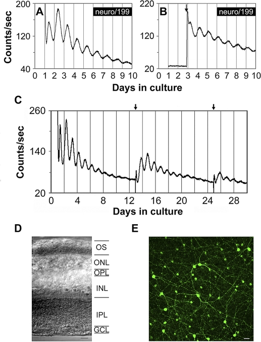
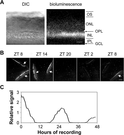
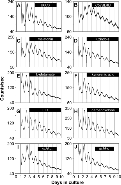
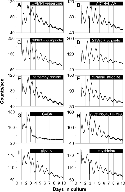
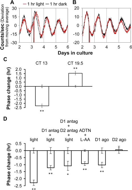
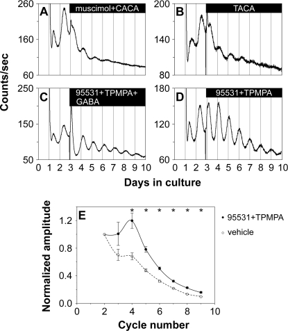
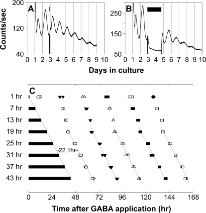
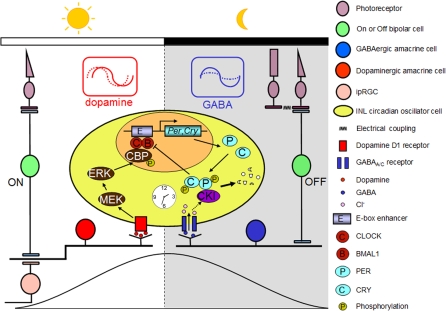
The influence of the mammalian retinal circadian clock on retinal physiology and function is widely recognized, yet the cellular elements and neural regulation of retinal circadian pacemaking remain unclear due to the challenge of long-term culture of adult mammalian retina and the lack of an ideal experimental measure of the retinal circadian clock. In the current study, we developed a protocol for long-term culture of intact mouse retinas, which allows retinal circadian rhythms to be monitored in real time as luminescence rhythms from a PERIOD2::LUCIFERASE (PER2::LUC) clock gene reporter. With this in vitro assay, we studied the characteristics and location within the retina of circadian PER2::LUC rhythms, the influence of major retinal neurotransmitters, and the resetting of the retinal circadian clock by light. Retinal PER2::LUC rhythms were routinely measured from whole-mount retinal explants for 10 d and for up to 30 d. Imaging of vertical retinal slices demonstrated that the rhythmic luminescence signals were concentrated in the inner nuclear layer. Interruption of cell communication via the major neurotransmitter systems of photoreceptors and ganglion cells (melatonin and glutamate) and the inner nuclear layer (dopamine, acetylcholine, GABA, glycine, and glutamate) did not disrupt generation of retinal circadian PER2::LUC rhythms, nor did interruption of intercellular communication through sodium-dependent action potentials or connexin 36 (cx36)-containing gap junctions, indicating that PER2::LUC rhythms generation in the inner nuclear layer is likely cell autonomous. However, dopamine, acting through D1 receptors, and GABA, acting through membrane hyperpolarization and casein kinase, set the phase and amplitude of retinal PER2::LUC rhythms, respectively. Light pulses reset the phase of the in vitro retinal oscillator and dopamine D1 receptor antagonists attenuated these phase shifts. Thus, dopamine and GABA act at the molecular level of PER proteins to play key roles in the organization of the retinal circadian clock.


Conflict of interest statement

Competing interests. The authors have declared that no competing interests exist.

Figures

Figure 1
Figure 1. Bioluminescence Rhythms from mPer2Luc Mouse Retinal Explants
(A) A representative PER2::LUC bioluminescence trace recorded from a B6C3 mPer2Luc mouse retinal explant. (B) A representative PER2::LUC bioluminescence trace recorded from a mouse retinal explant in which luciferin substrate was absent from the medium in the first two cycles. Arrow indicates addition of luciferin. (C) Long-term culture of an intact mouse retinal explant showing persistent circadian rhythms in PER2::LUC expression. Arrows indicate times of media changes. (D) Representative DIC image of vertical retinal sections. Retinal explants were cultured for 9 d in vitro (DIV), and vertical retinal slices were cut with a tissue slicer at 150 μm. GCL, ganglion cell layer; INL, inner nuclear layer; IPL, inner plexiform layer; ONL, outer nuclear layer; OPL, outer plexiform layer; OS, photoreceptor outer segments. Scale bar represents 20 μm. (E) Flat-mount view showing tyrosine hydroxylase immunoreactivity in retinal explants that were cultured for 9 DIV. The immunoreactive cells with relatively large somata and two to three thick primary processes that arise from the cell body are dopaminergic amacrine cells, whereas the immunoreactive cells with relatively small cell body and very few processes are type 2 catecholamine amacrine cells. Scale bar represents 20 μm.
Figure 2
Figure 2. Circadian Oscillation of PER2::LUC Bioluminescence in the Inner Nuclear Layer
(A) DIC image (left panel) and luminescence image (right panel) of a vertical retinal slice. Retinal slices were prepared around ZT 4, incubated in CO2 incubator for 1 d, and then imaged. Note that bioluminescence signals were primarily located in the inner nuclear layer. GCL, ganglion cell layer; INL, inner nuclear layer; IPL, inner plexiform layer; ONL, outer nuclear layer; OPL, outer plexiform layer; OS, photoreceptor outer segments. Scale bar represents 50 μm. (B) PER2::LUC bioluminescence in the inner nuclear layer of cultured vertical retinal slices showed a circadian variation with a peak at projected ZT 8–14. Shown are representative images acquired by 30-min exposures at 6-h intervals. Arrows point to membrane filters that became visible when signals in the inner nuclear layer were high. Scale bar represents 100 μm. (C) Continuous recording of PER2::LUC bioluminescence in a representative cultured vertical retinal slice. In this experiment, retinal slices were prepared around ZT 4, incubated in CO2 incubator for 2 d, and then images (30-min exposure) were continuously collected for 47 h, starting at projected ZT 9.
Figure 3
Figure 3. Retinal PER2::LUC Rhythms Do Not Require Communication via Melatonin, Glutamate, Sodium-Dependent Action Potentials or cx36-Containing Gap Junctions
(A) mPer2Luc retinal explants cultured from B6C3 mice exhibited sustained circadian rhythms in PER2::LUC expression. (B) mPer2Luc retinal explants cultured from C57BL/6J mice also showed robust circadian oscillation of PER2::LUC expression. (C–H) Circadian PER2::LUC expression rhythms persisted upon continuous application of: (C) melatonin (10 nM); (D) the melatonin MT1 receptor antagonist luzindole (5 μM); (E) l-glutamate (1 mM); (F) the broad-spectrum glutamate receptor antagonist kynurenic acid (0.5 mM); (G) the voltage-gated sodium channel blocker TTX (1 μM); and (H) the gap junction block carbenoxolone (100 μM). (I and J) Retinal explants cultured from cx36−/− mice (I) and cx36+/− mice (J) did not show differences in the damping rate of PER2::LUC rhythms. Bars indicate the duration of treatment.
Figure 4
Figure 4. Retinal PER2::LUC Rhythms Are Not Dependent on Communication via Dopamine, Acetylcholine, GABA, or Glycine
(A–F) Retinal circadian PER2::LUC expression rhythms persisted upon continuous application of: (A) the TH inhibitor L-AMPT (100 μM) along with the vesicular dopamine uptake inhibitor reserpine (10 μM); (B) the dopamine receptor agonist ADTN (100 μM), in the presence of L-AA (100 μM); (C) the dopamine D1 receptor agonist SKF-38393 (50 μM) along with the D2/D4 receptor agonist quinpirole hydrochloride (50 μM); (D) the dopamine D1 receptor antagonist SCH-23390 (50 μM) along with the D2 receptor antagonist sulpiride (50 μM); (E) the nonselective cholinergic agonist carbamoylcholine chloride (100 μM); and (F) the nicotinic acetylcholine receptor antagonist (+)-tubocurarine (100 μM) along with the muscarinic acetylcholine receptor antagonist atropine (100 μM). (G) GABA (1 mM) greatly suppressed the amplitude of retinal PER2::LUC rhythms. (H) Retinal PER2::LUC rhythms persisted during blockade of GABA receptors with the GABAA receptor antagonist SR-95531 (40 μM) along with the GABAB receptor antagonist CGP-35348 (100 μM) and the GABAC receptor antagonist TPMPA (100 μM). (I and J) Retinal PER2::LUC rhythms persisted upon continuous application of: (I) glycine (3 mM); and (J) the glycine receptor antagonist strychnine hydrochloride (50 μM). Bars indicate the duration of treatment.
Figure 5
Figure 5. Dopamine Shifts the Phase of Retinal PER2::LUC Rhythms
(A) Application of ADTN (100 μM) along with L-AA (100 μM) beginning at CT 3 advanced PER2::LUC rhythms compared to L-AA (100 μM) application alone. (B) Application of the D1 receptor agonist SKF 38393 (50 μM) beginning at CT 18 delayed PER2::LUC rhythms compared to vehicle (H2O). Data shown in (A) and (B) were baseline corrected by calculating a 24-h moving average of the raw data, and then the deviation from the moving average was plotted as a function of days in culture. Bars indicate the duration of treatment. (C) Phase change following application of dopamine agonists. Briefly, the peak times (as determined by ClockLab software) on the third cycle (after treatment) were subtracted from the peak times on the second cycle (before treatment) for both drug and vehicle, and then used to calculate the phase change of drug versus vehicle controls. Bars show drug phase changes (means ± SEM), error bars from x axis show ±SEM for vehicle controls in each group. Drug concentrations were as follows: ADTN, 100 μM; the D1 agonist SKF 38393, 50 μM; and the D2 agonist quinpirole, 50 μM. Double asterisks (**) indicate p < 0.01, Student's t-test; n = at least 4 for each drug treatment and for vehicle controls. (D) Lack of phase change following application of melatonin reagents. Phase change was calculated as in (C). Drug concentrations are indicated above columns. Vehicles used for 10 nM melatonin (mel), 10 μM melatonin, and 5 μM luzindole (luz) were 1 μl of 0.01% ethanol, 1 μl of 10% ethanol, and 1 μl of DMSO, respectively; n = at least 4 for each drug treatment and for vehicle controls. No significant effects were observed (p > 0.05 for all).
Figure 6
Figure 6. Light Resetting of the Phase of Retinal PER2::LUC Rhythms Is Mediated In Part through Dopamine D1 Receptors
(A and B) Phase changes of retinal PER2::LUC rhythms induced by 1-h light pulses beginning at CT 13 (A) or CT 19.5 (B). Red traces show rhythms from explants that received light; black traces are from paired controls that were handled similarly, but did not receive light. (C) Average data of light-induced phase shifts. Error bars from x-axis show ±SEM for controls in which retinal explant cultures were taken out from the LumiCycle and kept in darkness at 37 °C for 1 h. (D) Phase changes of retinal PER2::LUC rhythms. Retinal explant cultures were subjected to or treated with: column 1, 1-h light pulses (a reproduction from [C]); column 2, pretreatment with the D1 receptor antagonist SCH-23390 (D1 antag; 50 μM) for 15 min followed by 1-h light pulses; column 3, pretreatment with SCH-23390 and the D2 antagonist sulpiride (D2 antag; 50 μM) together for 15 min followed by 1-h light pulses; column 4, the nonselective dopamine receptor agonist ADTN (100 μM; in vehicle containing 100 μM L-AA); column 5, the D1 agonist SKF 38393 (D1 ago; 50 μM); and column 6,the D2 agonist quinpirole (D2 ago; 50 μM). The 1-h light pulses or dopaminergic treatments were initiated at CT 13. Phase change was calculated as in Figure 5. A single asterisk (*) indicates p < 0.05; double asterisks (**) indicate p < 0.01; Student's t-test; n = at least 3 for each treatment and for controls.
Figure 7
Figure 7. GABA Sets the Amplitude of Retinal PER2::LUC Rhythms in a Dose-Dependent Manner
(A–D) Representative PER2::LUC bioluminescence traces recorded from cultured mPer2Luc retinal explants that received different doses of GABA treatment. Bars indicate the duration of GABA treatment. (E) Dose-dependent inhibitory action of GABA on the amplitude of retinal PER2::LUC rhythms. The ratio of the peak-to-trough amplitude of the fourth cycle to that of the second cycle (A4/A2) was plotted as a function of the external GABA concentration. Data are represented as mean ± SEM (n = at least 3 explants per point).
Figure 8
Figure 8. GABA Acts through GABAA and GABAC Receptors
(A) Coapplication of the GABAA receptor agonist muscimol (200 μM) and the GABAC receptor agonist CACA (50 μM) to retinal explants mimicked the inhibitory action of 1 mM GABA on retinal PER2::LUC rhythms. (B) The GABAA and GABAC receptor agonist TACA (80 μM) inhibited PER2::LUC expression rhythms in a way similar to 1 mM GABA. (C) The GABAA receptor antagonist SR-95531 (40 μM) and the GABAC receptor antagonist TPMPA (100 μM) greatly attenuated the action of GABA when they were coapplied with 1 mM GABA. (D) Coapplication of SR-95531 (40 μM) and TPMPA (100 μM) increased the amplitude of retinal PER2::LUC rhythms. Bars in (A–D) indicate the duration of treatment. (E) Blockade of GABAA and GABAC receptors with 40 μM SR-95531 and 100 μM TPMPA (solid line, filled circles) significantly increased the amplitude of retinal PER2::LUC rhythms compared with vehicle-treated explants (dashed line, open circles). Mean peak-to-trough amplitudes (±SEM, n = 5; normalized to the amplitude of the second circadian cycle) of retinal PER2::LUC rhythms were plotted as a function of PER2::LUC cycle number in the LumiCycle. A single asterisk (*) indicates p < 0.007, post hoc t-test with Bonferroni-corrected α = 0.007.
Figure 9
Figure 9. Prolonged Application of GABA Stops the Retinal Clock
(A and B) Representative retinal explant cultures that received 1 h (A) or 43 h (B) of 3 mM GABA treatment. GABA treatment was started at the beginning of the third circadian cycle. Bars indicate the duration of GABA treatment. Treatment was terminated by fresh media change. (C) Shown are PER2::LUC expression peaks following GABA washout. Four retinal explants were sampled for each duration of GABA treatment. Bars indicate the duration of GABA treatment. Open circles, filled triangles, open triangles, filled squares, open squares, and filled diamonds indicate first, second, third, fourth, fifth, and sixth peak times, respectively, following GABA washout. Time 0 corresponds to the start of GABA treatment. In the samples with 19–43 h of GABA application, the first peaks appeared ca. 22 h after media change, and the reinitiated rhythms were phase locked to the termination of the GABA pulse, indicating that prolonged application of high dose of GABA stops retinal ensemble PER2::LUC rhythms.
Figure 10
Figure 10. GABA Acts Posttranscriptionally through Membrane Polarization and Casein Kinase
(A) Shown are relative mRNA abundance of Per1, Per2, Clock, and Bmal1 in retinal explants 8 h after treatment with 3 mM GABA (black) or vehicle (grey) started at the beginning of the third circadian cycle. Clock gene mRNA levels were normalized to the mRNA levels of GAPDH. The average mRNA levels of vehicle-treated samples were set to 1.0. Data are presented as means ± SEM (n = 4). GABA did not significantly change the transcript levels of Per2 and Clock, while significantly increased the transcript levels of Per1 and Bmal1. A single asterisk (*)indicates p < 0.05; double asterisks (**) indicate p < 0.01; Student's t-test. (B) GABA (1 mM) greatly suppressed retinal PER2::LUC rhythms. (C) Depolarization with elevated K+ (4 mM) partially blocked the inhibitory effect of GABA (1 mM) on retinal PER2::LUC rhythms. (D) Prolonged application of KCl (4 mM) modestly increased rhythmic PER2::LUC amplitudes. (E) Inhibition of casein kinase activity with CKI-7 (50 μM) partially rescued the inhibitory action of 1 mM GABA on retinal PER2::LUC rhythms. (F) KCl (4 mM) and CKI-7 (50 μM) greatly rescued the inhibitory action of GABA (1 mM) on retinal PER2::LUC rhythms when they were co-applied with GABA. (G) When CKI-7 (50 μM) was applied alone, it lengthened the period of retinal PER2::LUC rhythms. Bars in (B–G) indicate the duration of treatment.
Figure 11
Figure 11. Model for the Circadian Clock Organization in the Mouse Retina
Dopaminergic amacrine cells (red) and GABAergic cells (blue) reinforce the autonomously generated “day” and “night” states of oscillator cells (yellow) in the inner nuclear layer (INL) through rhythmic secretion of dopamine and GABA, respectively. Dopamine transmission stimulated by excitatory light input from M-cones via ON bipolar cells and/or from intrinsically photosensitive retinal ganglion cells (ipRGCs) likely acts to phase-shift retinal circadian oscillators and reinforce the rising phase of the “day” state of the retinal clock. At night (i.e., molecularly, roughly the falling phase of PER protein rhythms), excitatory input from OFF bipolar cells enhances the activity of GABA, which suppresses rhythmic amplitude of PER oscillations through the GABAA and GABAC receptors and facilitates the molecular resetting effects of dopamine. In addition, endogenous GABA is proposed to reinforce the falling phase of PER rhythms through fostering degradation of accumulated PER protein.

References

    1. LaVail MM. Rod outer segment disk shedding in rat retina: relationship to cyclic lighting. Science. 1976;194:1071–1074. - PubMed
    1. Grace MS, Wang LM, Pickard GE, Besharse JC, Menaker M. The tau mutation shortens the period of rhythmic photoreceptor outer segment disk shedding in the hamster. Brain Res. 1996;735:93–100. - PubMed
    1. LaVail MM. Circadian nature of rod outer segment disc shedding in the rat. Invest Ophthalmol Vis Sci. 1980;19:407–411. - PubMed
    1. Grace MS, Chiba A, Menaker M. Circadian control of photoreceptor outer segment membrane turnover in mice genetically incapable of melatonin synthesis. Vis Neurosci. 1999;16:909–918. - PubMed
    1. Li L, Dowling JE. Zebrafish visual sensitivity is regulated by a circadian clock. Vis Neurosci. 1998;15:851–857. - PubMed

Publication types

MeSH terms