. 2009 Jan-Feb;40(1):9.
doi: 10.1051/vetres:2008047. Epub 2008 Oct 30.

Intermittent Escherichia coli O157:H7 colonisation at the terminal rectum mucosa of conventionally-reared lambs

Affiliations

Intermittent Escherichia coli O157:H7 colonisation at the terminal rectum mucosa of conventionally-reared lambs

Angus Best et al. Vet Res. 2009 Jan-Feb.

Abstract

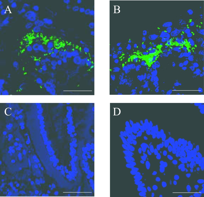
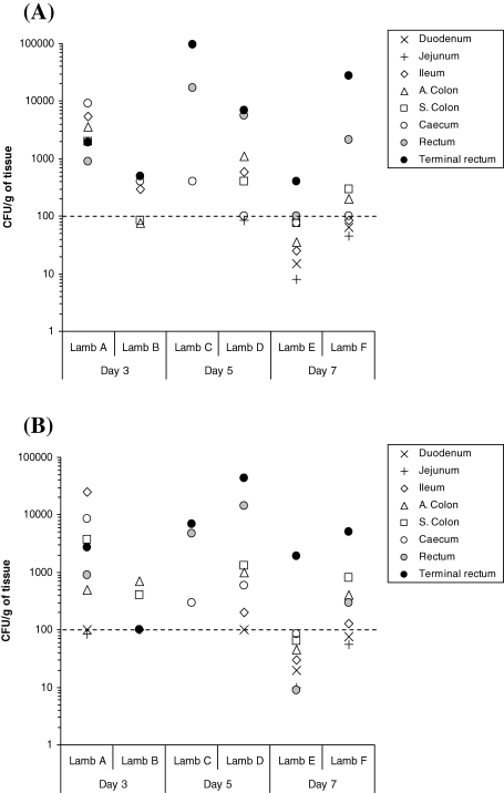
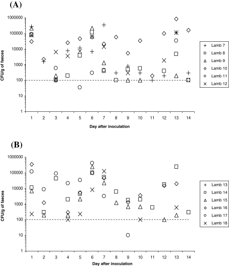
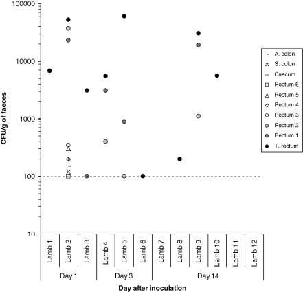
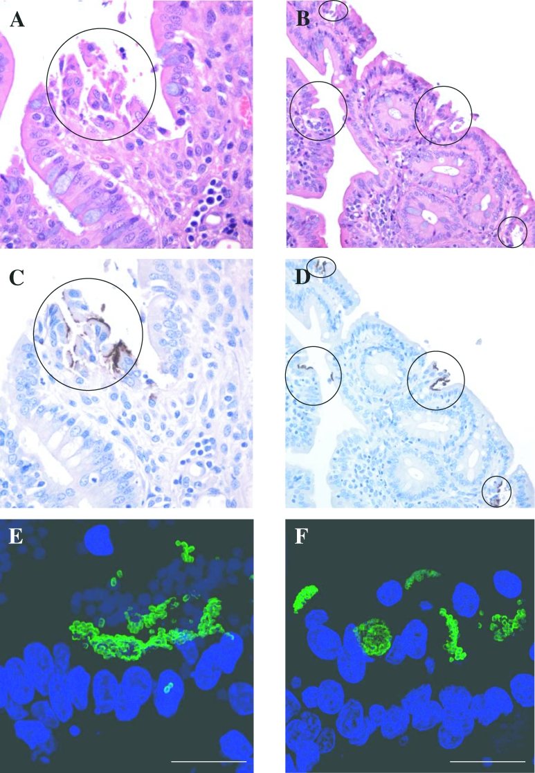
In cattle, the lymphoid rich regions of the rectal-anal mucosa at the terminal rectum are the preferred site for Escherichia coli O157:H7 colonisation. All cattle infected by rectal swab administration demonstrate long-term E. coli O157:H7 colonisation, whereas orally challenged cattle do not demonstrate long-term E. coli O157:H7 colonisation in all animals. Oral, but not rectal challenge of sheep with E. coli O157:H7 has been reported, but an exact site for colonisation in sheep is unknown. To determine if E. coli O157:H7 can effectively colonise the ovine terminal rectum, in vitro organ culture (IVOC) was initiated. Albeit sparsely, large, densely packed E. coli O157:H7 micro-colonies were observed on the mucosa of ovine and control bovine terminal rectum explants. After necropsy of orally inoculated lambs, bacterial enumeration of the proximal and distal gastrointestinal tract did suggest a preference for E. coli O157:H7 colonisation at the ovine terminal rectum, albeit for both lymphoid rich and non-lymphoid sites. As reported for cattle, rectal inoculation studies were then conducted to determine if all lambs would demonstrate persistent colonisation at the terminal rectum. After necropsy of E. coli O157:H7 rectally inoculated lambs, most animals were not colonised at gastrointestinal sites proximal to the rectum, however, large densely packed micro-colonies of E. coli O157:H7 were observed on the ovine terminal rectum mucosa. Nevertheless, at the end point of the study (day 14), only one lamb had E. coli O157:H7 micro-colonies associated with the terminal rectum mucosa. A comparison of E. coli O157:H7 shedding yielded a similar pattern of persistence between rectally and orally inoculated lambs. The inability of E. coli O157:H7 to effectively colonise the terminal rectum mucosa of all rectally inoculated sheep in the long term, suggests that E. coli O157:H7 may colonise this site, but less effectively than reported previously for cattle.


Figures

Figure 1.
Figure 1.
Observation of E. coli O157:H7 densely packed micro-colonies on ovine (A) and bovine (B) terminal rectum explants as visualised by confocal microscopy. Dapi (blue) = nuclei. FITC (green) = E.coli O157. (C) and (D) are uninfected ovine and bovine terminal rectum explants, respectively. E. coli O157:H7 was incubated with explants for 8 h with changing of the media occurring every hour after the first 2 h. Bar (A) = 32.17 μm, (B) = 27.5 μm, C = 49.26 μm, D = 31.77 μm.
Figure 2.
Figure 2.
Evaluation of E. coli O157:H7 tropisms to lymphoid (A) and non-lymphoid (B) regions of the GIT in young lambs orally inoculated with approximately 1 × 1010 CFU, as determined by bacterial enumeration after necropsy. All data points on or above the dotted line were obtained after direct plating. All data points below the dotted line were obtained after enrichment of faecal samples negative for growth after direct plating. These enrichment data points are only nominal and could represent any value between 1 and 99 CFU/g faeces−1. Samples where no E. coli O157:H7 growth was detected after enrichment are not shown. N.B.: Non-lymphoid ileum was actually sampled from a part of the ileum where less dense lymphoid tissue reside, as the majority of ovine ileum exhibits lymphoid rich sites.
Figure 3.
Figure 3.
The distribution of E. coli O157:H7 shedding over time after the inoculation of 6-week-old lambs either (A) rectally or (B) orally with approximately 1 × 1010 CFU. All data points shown are from direct plating only. E. coli O157:H7 was not detected from the faeces of lamb 21 during the fourteen day sampling period. Samples where no E. coli O157:H7 growth was detected after enrichment are not shown.
Figure 4.
Figure 4.
Recovery of E. coli O157:H7 from the gastrointestinal tract of young lambs rectally inoculated with approximately 1 × 1010 CFU. All data points shown on or above the dotted line are from direct plating only. No E. coli O157:H7 growth was detected after enrichment and zero values are not shown.
Figure 5.
Figure 5.
Light and confocal microscopy of E. coli O157:H7 localised adherence and micro-colony formation at the terminal rectum mucosa of rectally inoculated young lambs at day 3 (Lamb 5: A, C and E) and 14 (Lamb 9: B, D and F) after inoculation. Open circles denote localised adherence. Staining: (A and B) = Haematoxylin and Eosin; (C and D) = Meyer’s haematoxylin (terminal rectum-blue), immunoperoxidase (E. coli O157-brown); (E and F) Dapi (blue) = nuclei; FITC (green) =  E.coli O157. Magnification (AandC) = 400×, (B and D) = 200×. Bar (E) = 20 μm, (F) = 18 μm.

References

    1. Best A., La Ragione R.M., Clifford D., Cooley W.A., Sayers A.R., Woodward M.J.. A comparison of Shiga-toxin negative Escherichia coli O157 aflagellate and intimin deficient mutants in porcine in vitro and in vivo models of infection. Vet. Microbiol. 2006;113:63–72. - PubMed
    1. Borczyk A.A., Karmali M.A., Lior H., Duncan L.M.. Bovine reservoir for verotoxin-producing Escherichia coli O157:H7. Lancet. 1987;1:98. - PubMed
    1. Chapman P.A., Cerdan-Maló A.T., Ellin M., Ashton R.. Escherichia, coli O157 in cattle and sheep at slaughter, on beef and lamb carcasses and in raw beef and lamb products in South Yorkshire, UK. Int. J. Food Microbiol. 2001;64:139–150. - PubMed
    1. Chase-Topping M.E., McKendrick I.J., Pearce M.C., MacDonald P., Mathews L., Halliday J.. et al. Risks factors for the presence of high-level shedding of Escherichia coli O157 on Scottish farms. J. Clin. Microbiol. 2007;45:1594–1603. - PMC - PubMed
    1. Cobbold R.N., Hancock D.D., Rice D.H., Berg J., Stilborn R., Hovde C.J., Besser T.E.. Rectoanal junction colonisation of feedlot cattle by Escherichia coli O157:H7 and its association with supershedders and excretion dynamics. Appl. Environ. Microbiol. 2007;73:1563–1568. - PMC - PubMed

Publication types