. 2009 Feb;46(5):830-9.
doi: 10.1016/j.molimm.2008.09.018. Epub 2008 Oct 29.

Short leucine-rich glycoproteins of the extracellular matrix display diverse patterns of complement interaction and activation

Affiliations

Short leucine-rich glycoproteins of the extracellular matrix display diverse patterns of complement interaction and activation

Andreas P Sjöberg et al. Mol Immunol. 2009 Feb.

Abstract

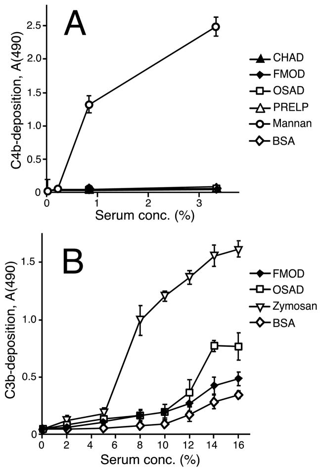
The extracellular matrix consists of structural macromolecules and other proteins with regulatory functions. An important family of the latter class of molecules found in most tissues is the small leucine-rich repeat proteins (SLRPs). We have previously shown that the SLRP fibromodulin binds directly to C1q and activates the classical pathway of complement. In the present study we further examine the interactions between SLRPs and complement. Osteoadherin, like fibromodulin, binds C1q and activates the classical pathway strongly while moderate activation is seen in the terminal pathway. This can be explained by the interaction of fibromodulin and osteoadherin with factor H, a major soluble inhibitor of complement. Also, chondroadherin was found to bind C1q and activate complement, albeit to a lesser extent. Chondroadherin also binds factor H. We confirm published data showing that biglycan and decorin bind C1q but do not activate complement. In this study a similar pattern is seen for lumican although its affinity for C1q is lower than for biglycan and decorin. Furthermore, using electron microscopy and radiolabeled SLRPs, we demonstrate two different classes of SLRP binding sites on C1q, to head and stalk respectively, where only binding to the head appears to be activating. We propose a role for SLRPs in the regulation of complement activation in diseases involving the extracellular matrix, particularly those characterized by chronic inflammation such as rheumatoid arthritis, atherosclerosis, osteoarthritis and chronic obstructive lung disease.


Figures

Fig. 1
Fig. 1. Structural overview of the cartilage related SLRPs used in this study
All proteins contain an LRR-region, which in all but one case (CHAD) includes varying numbers of glycosylation substitution sites. Asterisks indicate tyrosine sulfation sites and + indicates positively charged arginine- or lysine rich patches. CS: chondroitin sulfate, DS: dermatan sulfate, KS: keratan sulfate.
Fig. 2
Fig. 2. Abilities of SLRPs to activate the complement system
SLRPs and controls were coated onto microtiter plate wells over night followed by blocking of the plate to prevent further protein binding to the plastic surface. Increasing concentrations of normal human serum (NHS) were added and incubated at 37°C. Specific antibodies were used to detect deposition of complement components indicating complement activation. The pattern of C1 (A, B), C4b (C, D), C3b (E, F) and C9 (G, H) deposition is plotted in the figure. The data were collected from two representative duplicate experiments (n=4). Error bars show standard deviation. Statistics were performed for all data (two-tailed Student’s T-test): In panels A & B, all points above 0.1 % NHS were significantly different (p < 0.01) from BSA except for one point each on decorin (highest NHS concentration) and lumican (second highest NHS concentration). In C & D, significance was seen for all NHS concentrations on FMOD & OSAD (p < 0.001) and for the highest concentration on PRELP (p < 0.001). In E & F, all data points for FMOD and OSAD showed significance (p < 0.001 and p < 0.01, respectively) while calculations for only the highest serum concentration point on CHAD, lumican and PRELP showed significance (p < 0.05 for the former two and p < 0.001 for the latter). In G & H, significance was seen for all data points on FMOD (p < 0.05) and OSAD (p < 0.001) as well as the highest concentration points on CHAD and PRELP (p < 0.01).
Fig. 3
Fig. 3. Abilities of SLRPs to induce complement activation via the lectin and alternative pathways
SLRPs and controls were coated onto microtiter plate wells then the plates were blocked for further direct binding. Human serum diluted to various concentrations was incubated at 37°C. Specific antibodies against C3b and C4b were added to detect complement activation on the surfaces. In A, the lectin pathway was assayed by using C1q-deficient serum, which eliminates classical pathway contribution. Mannan was used as the positive control for C4b-deposition via the lectin pathway. B shows complement activation as C3b-deposition from NHS in a buffer specific for the alternative pathway. Here, zymosan was used as the positive control. In both panels the data are from two representative duplicate experiments (n=4). Error bars show standard deviation. In B the differences between activation on both FMOD & OSAD and BSA are statistically significant for all serum dilutions above 5 % (p < 0.05; two tailed Student’s T-test).
Fig. 4
Fig. 4. Binding of C1q to SLRPs
A, B Plates were coated with SLRPs or controls, blocked and then incubated with C1q at the concentrations indicated. Bound C1q was detected using specific polyclonal antibodies. The data were collected from two representative duplicate experiments (n=4). Error bars denote standard deviation. The difference between C1q binding on SLRPs and BSA is statistically significant (p < 0.01; two-tailed Student’s T-test) for all data points above 0 μg/ml C1q except for the two lowest C1q concentrations on PRELP. C C1q was injected at least twice at increasing concentrations (6.5 nM–416 nM) over the Biacore CM5-chip with bound OSAD and in parallel over a control flow cell that was used to subtract nonspecific signal. The amount of bound C1q was measured in arbitrary response units (RU, inset). The response obtained for each concentration of C1q at equilibrium (Req) was plotted against concentration of C1q, which allowed for estimation of KD presented in Table 1.
Fig. 5
Fig. 5. Binding of SLRPs to intact and truncated C1q species and ion strength dependence of the interactions
A Wells of microtiter plates were coated with intact C1q, C1q head, C1q tail or BSA, blocked and then incubated with the 125I-labeled SLRPs indicated (50,000 counts per well). After washing, the numbers of counts retained were determined and values are expressed as the proportion of counts added. For each SLRP, the data were normalized against the average number of counts retained for intact C1q. The displayed data represent the mean of quadruplicate determinations (n=4), with error bars representing the standard deviation. Asterisks indicate significance (two-tailed Student’s T-test) accordingly: *, p < 0.05; ***, p < 0.001, ns - not significant. B SLRPs were coated as in the direct C1q-binding assay and C1q was added at 5 μg/ml in buffers with varying ionic strength or with 150 mM NaCl and 5 mM EDTA. Bound C1q was detected with specific antibodies. The wells for each SLRP were developed separately to allow for proper comparison between the different buffer conditions. The data were collected from two representative duplicate experiments (n=4) and error bars show standard deviation. In B all changes, as compared to the respective levels for 150 mM, are statistically significant except for the effect of EDTA on C1q binding to biglycan and decorin (two-tailed Student’s T-test).
Fig. 6
Fig. 6. Visualization of SLRPs binding to C1q using electron microscopy
The complexes between C1q and gold-labeled SLRPs were visualized by negative staining. A Single molecules of C1q without any ligand. B Single molecules of C1q with one to three bound gold-labeled OSAD molecules. C Single molecules of C1q with one to three bound gold-labeled CHAD molecules. D Single molecules of C1q with one to four bound gold-labeled decorin molecules. E Single molecules of C1q with one to four bound gold-labeled biglycan molecules. F Single molecules of C1q with one to four bound gold-labeled lumican molecules. Ninety to 95 % of all interactions occurred according to the patterns presented (n ≥ 300 for each respective interaction). Scale bar is 25 nm.
Fig. 7
Fig. 7. Interactions between SLRPs and FH and the effect of FH on C9-deposition on SLRPs
A The indicated SLRPs were immobilized in microtiter plates, the wells were blocked and incubated with two concentrations of full-length human FH. The amount of bound FH was assessed with a specific antibody. B CHAD was coated onto microtiter plates, and after blocking, either full length FH or one of two constructs representing polymorphic variants of CCP domains 6–8 of FH was allowed to bind. Bound protein was detected with a specific polyclonal antibody. In A the data were collected from two representative duplicate experiments (n=4), while B shows data from a representative experiment done in triplicate (n=3). Error bars denote standard deviation. Asterisks indicate significance accordingly: **, p < 0.01; ***, p < 0.001.

References

    1. Antonsson P, Heinegård D, Oldberg A. Posttranslational modifications of fibromodulin. J Biol Chem. 1991;266:16859–61. - PubMed
    1. Aronen M, Lehto T, Meri S. Regulation of alternative pathway complement activation by an interaction of C-reactive protein with factor H. Immunology and Infectious Diseases. 1992;3:83–87.
    1. Baschong W, Lucocq JM, Roth J. “Thiocyanate gold”: small (2–3 nm) colloidal gold for affinity cytochemical labeling in electron microscopy. Histochemistry. 1985;83:409–11. - PubMed
    1. Bing DH, Almeda S, Isliker H, Lahav J, Hynes RO. Fibronectin binds to the C1q component of complement. PNAS. 1982;79:4198–4201. - PMC - PubMed
    1. Blom AM, Kask L, Dahlbäck B. CCP1–4 of the C4b-binding protein α-chain are required for Factor I mediated cleavage of C3b. Mol Immunol. 2003a;39:547–56. - PubMed

Publication types

MeSH terms