Review
. 2009 Jan;130(1):7-15.
doi: 10.1016/j.clim.2008.08.015. Epub 2008 Oct 28.

Gene-specific control of the TLR-induced inflammatory response

Affiliations
Review

Gene-specific control of the TLR-induced inflammatory response

Simmie L Foster et al. Clin Immunol. 2009 Jan.

Abstract

Toll-like receptors (TLRs) induce a complex inflammatory response that functions to alert the body to infection, neutralize pathogens, and repair damaged tissues. An excessive or persistent inflammatory response can be fatal, so multiple regulatory mechanisms have evolved to control the extent and duration of inflammation. Our current understanding of the control of inflammation is based on negative regulation of TLR signaling. However, TLR-induced genes have diverse functions, and control of signaling pathways does not allow for groups of genes with distinct functions to be differentially regulated. Recent evidence suggests that many inflammatory genes are instead regulated by epigenetic modifications to individual promoters. This level of control allows a single gene to be expressed or silenced according to its function, irrespective of other genes induced by the same receptor, and therefore is "gene-specific." Gene-specific control of the TLR-induced inflammatory response is an emerging paradigm in the study of inflammation, and may provide the basis for selective modulation of the inflammatory response.


Figures
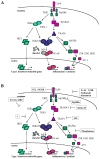
Figure 1
Figure 1
The TLR4 signaling pathway is highly regulated. A. Following LPS binding, TLR4 signals through two main adaptor molecules, MyD88 and TRIF. In the MyD88 dependent pathway, IRAK1 and IRAK4 are phosphorylated, then dissociate from the complex and activate TRAF6. TRAF6 activates the MAP kinases ERK, JNK, and p38, as well as the IKK complex. IKK phosphorylates IκBα, leading to its degradation, releasing NFκB to enter the nucleus and activate gene transcription. In the TRIF dependent pathway, TBK-1 phosphorylates IRF3, which then dimerizes and enters the nucleus to transcribe Type 1 IFN-inducible genes. TRIF signaling also leads to a delayed NFκB response. B. TLR4 induces multiple negative regulators. A20 deubiquitinates and deactivates TRAF6. IRAKM may inhibit dissociation of IRAK1 from the receptor complex. SIGRR and ST2 are inhibitory cell-surface receptors. SOCS1 inhibits IFNR signaling, SHIP antagonizes PI3K, and MyD88s is a non-functional splice variant of MyD88. An unknown regulator likely inhibits IRF3 phosphorylation. IL-10, TGFβ, and cholinergic stimulation also inhibit induction of proinflammatory cytokines. Schematic adapted with permission of D. Hargreaves.
Figure 2
Figure 2
The TLR4-induced inflammatory response has multiple components with distinct regulatory requirements. Upon recognizing bacterial infection, TLR4 signaling initiates the inflammatory response gene program. Inflammatory response genes fall into different functional groups, or components. Each component is regulated according to its role in the immune response and its potential for deleterious effects on the body if produced excessively. For example, proinflammatory cytokines and vasoactive factors (clotting factors, histamine) need only be induced transiently, and require tight regulation to prevent systemic shock. Antimicrobial proteins need to be produced for as long as pathogens persist, have no potential to cause shock, and are not highly regulated. For some components, such as oxidative killing, which have both direct antimicrobial activity and potential to damage tissues, the regulatory requirements are less clear, and must be tissue-type and context-dependent.
Figure 3
Figure 3
LPS tolerance is an example of gene-specific regulation of the inflammatory response. Following LPS stimulation of naïve macrophages, both class T and class NT promoters recruit transcription factors and display increased histone acetylation, histone H3K4 trimethylation, and accessibility (represented by yellow starburst). In tolerant macrophages stimulated with LPS, class T promoters are deacetylated and inaccessible. Class NT promoters, on the other hand, are inducibly reacetylated, remethylated, and made accessible with faster kinetics.

References

    1. Nathan C. Points of control in inflammation. Nature. 2002;420:846–52. - PubMed
    1. Karin M, Lawrence T, Nizet V. Innate immunity gone awry: linking microbial infections to chronic inflammation and cancer. Cell. 2006;124:823–35. - PubMed
    1. Takeda K, Kaisho T, Akira S. Toll-like receptors. Annu Rev Immunol. 2003;21:335–76. - PubMed
    1. Yeaman MR, Yount NY. Unifying themes in host defence effector polypeptides. Nat Rev Microbiol. 2007;5:727–40. - PubMed
    1. Berczi I. Neurohormonal host defense in endotoxin shock. Ann N Y Acad Sci. 1998;840:787–802. - PubMed

Publication types

MeSH terms

Substances