. 2009 Jan 15;18(2):328-40.
doi: 10.1093/hmg/ddn359. Epub 2008 Oct 29.

Twinkle mutations associated with autosomal dominant progressive external ophthalmoplegia lead to impaired helicase function and in vivo mtDNA replication stalling

Affiliations

Twinkle mutations associated with autosomal dominant progressive external ophthalmoplegia lead to impaired helicase function and in vivo mtDNA replication stalling

Steffi Goffart et al. Hum Mol Genet. .

Abstract

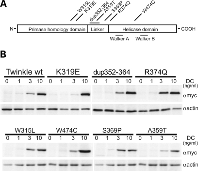
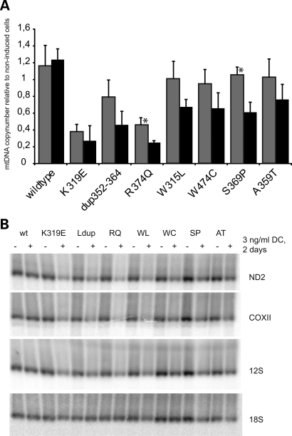
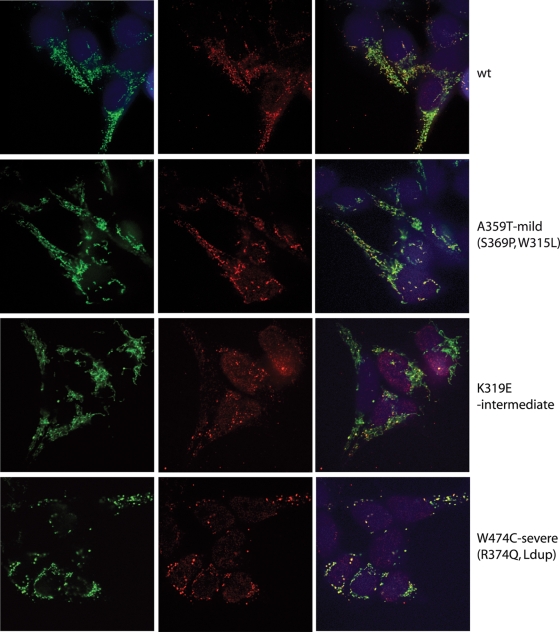
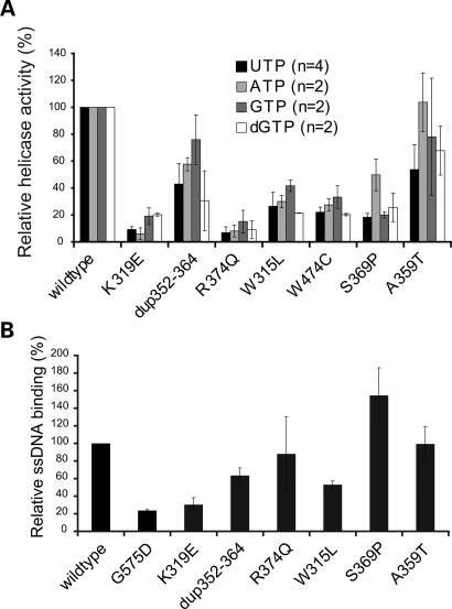
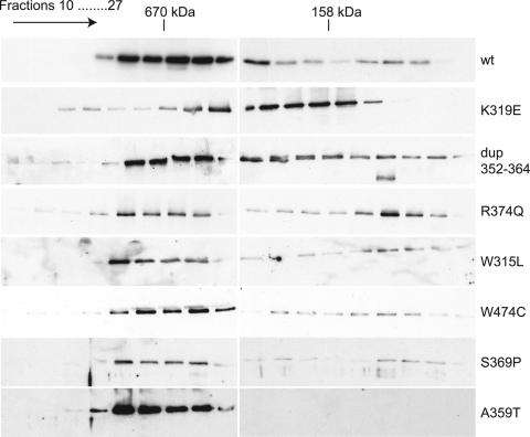
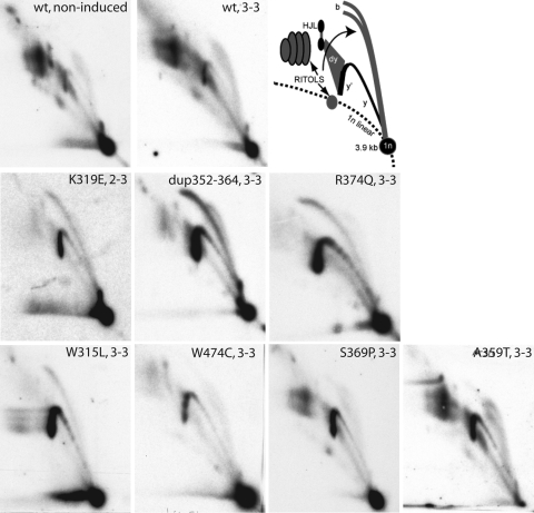
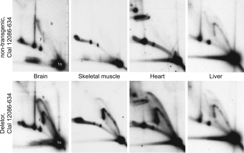
Mutations in the mitochondrial helicase Twinkle underlie autosomal dominant progressive external ophthalmoplegia (PEO), as well as recessively inherited infantile-onset spinocerebellar ataxia and rare forms of mitochondrial DNA (mtDNA) depletion syndrome. Familial PEO is typically associated with the occurrence of multiple mtDNA deletions, but the mechanism by which Twinkle dysfunction induces deletion formation has been under debate. Here we looked at the effects of Twinkle adPEO mutations in human cell culture and studied the mtDNA replication in the Deletor mouse model, which expresses a dominant PEO mutation in Twinkle and accumulates multiple mtDNA deletions during life. We show that expression of dominant Twinkle mutations results in the accumulation of mtDNA replication intermediates in cell culture. This indicated severe replication pausing or stalling and caused mtDNA depletion. A strongly enhanced accumulation of replication intermediates was evident also in six-week-old Deletor mice compared with wild-type littermates, even though mtDNA deletions accumulate in a late-onset fashion in this model. In addition, our results in cell culture pointed to a problem of transcription that preceded the mtDNA depletion phenotype and might be of relevance in adPEO pathophysiology. Finally, in vitro assays showed functional defects in the various Twinkle mutants and broadly agreed with the cell culture phenotypes such as the level of mtDNA depletion and the level of accumulation of replication intermediates. On the basis of our results we suggest that mtDNA replication pausing or stalling is the common consequence of Twinkle PEO mutations that predisposes to multiple deletion formation.


Figures

Figure 1.
Figure 1.
Expression of Twinkle adPEO mutations in HEK293 Flp-In™ T-REx™ cells. (A) Schematic presentation of the human Twinkle protein and localization of the adPEO-related mutations investigated in this study. Most mutations are located at the end of the N-terminal domain or in the linker region of Twinkle, only the W474C point mutation lies within the helicase domain. (B) Expression levels of adPEO Twinkle variants. Expression was induced for three days with the indicated amounts of doxycycline (DC) and protein expression was analyzed by western blot analysis against the myc-tagged Twinkle variants. At 1 ng/ml DC no increase in transgene expression could be observed, although mild effects on mtDNA replication were already detectable (data not shown). Maximal expression was reached with 10 ng/ml DC.
Figure 2.
Figure 2.
Mutant expression causes rapid mtDNA copynumber and transcript level depletion. (A) While overexpression of wild-type Twinkle transiently increased the relative copynumber of mtDNA per cell, expression of adPEO variants lead to rapid mtDNA depletion. The loss of mtDNA correlated approximately with the severity of the biochemical and replication defects measured in this study. MtDNA levels of cells induced with 3 ng/ml doxycycline for two days (grey bars) or three days (black bars) were quantified by duplex Taqman PCR and normalized to non-induced cells of the same cell line. Four to six independently isolated samples were measured in triplicate with the exception of the two samples indicated with an asterisk that were isolated twice and measured in triplicate. Similar results were obtained by Southern blot analysis (not shown). Error bars show the standard deviation. (B) Also the steady-state levels of mtDNA transcripts were decreased upon Twinkle mutant expression, while the overexpression of wild-type Twinkle and the resulting mtDNA increase did not change transcript levels. RNA from cells grown for two days with or without 3 ng/ml DC was analysed by Northen blot using probes against several mitochondrial transcripts as indicated and 18S rRNA. Note that the decrease in transcripts occurred in advance of mtDNA depletion (Fig. 2A). No effect on protein levels of representative, nuclear encoded, respiration complex subunits were detected by western blot analysis after 3 days (data not shown). Ldup refers to dup352–364.
Figure 3.
Figure 3.
Some adPEO Twinkle mutants alter the localization of the protein in mtDNA nucleoids. The subcellular localization of the Twinkle variants was determined by immunofluorescence. While wild-type Twinkle co-localized with mtDNA in punctate nucleoids within mitochondria, the disease variants K319E Twinkle variants localized only partially with nucleoids, a proportion of the protein was diffusely localized in the mitochondrial lumen, while the nucleoids often appeared enlarged (similar results were observed with the R374Q and dup 352–364 mutants; Supplementary Material, Fig. S2). The W474C variant caused aggregation of nucleoids without a more diffuse distribution of the protein, while the A359T mutation decreased the Twinkle localization in nucleoids without altering their size and number extensively. The effects of expression of the various mutants were categorized as mild, moderate and severe as indicated in the Figure. Cells were induced with 3 ng/ml doxycycline for 3 days, immunocytochemistry was performed using specific antibodies against the myc-tagged Twinkle variants (green) and DNA (red). Images were taken with the same settings for exposure, laser intensity, etc. and in addition were processed for brightness and contrast in the same manner. All mutants are shown in Supplementary Material, Fig. S2.
Figure 4.
Figure 4.
AdPEO mutations affect the unwinding and ssDNA binding activity of Twinkle. (A) Purified protein extracts of the various Twinkle mutations showed clearly reduced maximal unwinding activity in in vitro assays using an M13-based substrate. Most adPEO mutants had less than 25% remaining unwinding activity with UTP, while dup352–364 and A359T were around 50% active compared with wild-type protein (P < 0.05 for dup 352–364 and A359T, P < 0.01 for all others). Other nucleotides (ATP, GTP and dGTP) stimulated the unwinding activity of Twinkle to a lesser extend, but were giving approximately similar decreases for the mutants, only A359T exhibited full activity with ATP. (B) Twinkle ssDNA binding assays were performed essentially as described previously (30) (see also Materials and Methods). We used the G575D mutation previously described by us (22) as an additional control as this mutation was expected to have clearly reduced DNA affinity and indeed showed very poor binding even compared with the various adPEO Twinkle mutants. Of the adPEO mutants, K319E, dup352–364 and W315L showed reduced binding, whereas R374Q and A359T showed near normal ssDNA affinity. The S369P in our hands showed a moderately increased binding. Note that the W474C is not shown because this protein with several repeated isolations gave spurious results. Binding is expressed relative to the binding of the wild-type protein set at 100%.
Figure 5.
Figure 5.
Some adPEO mutations decrease the stability of Twinkle hexamers. Gel filtration chromatography of purified Twinkle proteins indicated the formation of hexameric complexes in both wild-type and mutant proteins. The same results were obtained in the presence or absence of Mg2+ ions and under salt conditions from 100 to 500 mm NaCl (data not shown). The partial delay and the extensive trailing during the chromatography run indicated the relative instability of the hexamers of K319E, R374Q and dup352–364. Following gel filtration all collected fractions were concentrated and analyzed by western blot analysis as detailed in Materials and Methods.
Figure 6.
Figure 6.
MtDNA replication is stalling upon Twinkle mutant expression. 2DNAGE analysis of mtDNA replication intermediates from cells expressing Twinkle variants for two to three days (2–3: two days of induction with 3 ng/ml doxycycline; 3–3: three days induction with 3 ng/ml doxycycline). 2DNAGE samples for all panels consisted of purified mtDNA digested with HincII and probed with a radiolabeled cytochrome b gene fragment (nt 14 846–15 357). The detected fragment includes the non-coding region of mtDNA, the cytochrome b, ND6, part of the ND5 gene and intervening tRNA genes (nt 13 636–1006). The upper two panels show a comparison of mtDNA replication in non-induced and Twinkle wild-type overexpressing cells and the interpretation based on earlier 2DNAGE analysis of mtDNA RIs (22). 1n, 3.9 kb non-replicating HincII fragment. b, bubble arcs. MtDNA bubble arcs are usually very sensitive to RNase H due to the presence of patches of RNA–DNA especially on the lagging strand; therefore a second, degraded bubble arc is visible in cells incorporating RNA into the lagging strand. y and y′ indicate ascending and descending parts of the y arc and (dy) indicates double-Y structures typical for the termination region of replication. These will eventually form resolution intermediates resembling Holliday junctions (HJL-Holliday junction like molecules). Other higher molecular weight fragments arise from the failure to cut RNA–DNA hybrids at a restriction site, these as well as the RNA-rich bubble arcs are marked as RITOLS (RNA Incorporation ThroughOut the Lagging Strand). Overexpression of wild-type Twinkle did not change the replication pattern, the only notable difference was a reproducible reduction in one of the HJL RIs as described earlier. In contrast the stronger disease mutations caused accumulation of replication intermediates on the y- and bubble arc (y and b) and a decrease in all RITOLS related features. The milder disease mutations S359P and A369T had only a moderately enhanced descending y-arc (y′) (note that the 1n spot in the A359T panel has run of the gel). The helicase domain mutation W474C was exceptional as no bubble arc, indicating initiation in the non-coding region, was clearly visible, while similar to other strong mutants RITOLS had almost completely disappeared. This was even more clearly visible after induction with 10 ng/ml DC for three days as is illustrated in Supplementary Material, Fig. S3.
Figure 7.
Figure 7.
Deletor mice expressing the mouse variant of dup352–364 Twinkle also show mtDNA replication stalling. 2DNAGE analysis of brain, skeletal muscle, heart and liver mtDNA from young Deletor mice expressing the mouse Twinkle mutation equivalent to the human dup352–364 Twinkle variant show, except for liver, a similar strong accumulation of replication intermediates on both y- and bubble arc as in cultured cells. MtDNA was isolated from 6-week-old transgenic mice and non-transgenic siblings and probed against the OH-containing ClaI fragment (nts 12086–634). A similar replication stalling as observed in brain was also observed in kidney (data not shown).

Similar articles

Cited by

References

    1. Schaefer A.M., Blakely E.L., He L., Whittaker R.G., Taylor R.W., Chinnery P.F., Turnbull D.M. Prevalence of mitochondrial DNA disease in adults. Ann. Neurol. 2008;63:35–39. - PubMed
    1. Majamaa K., Moilanen J.S., Uimonen S., Remes A.M., Salmela P.I., Karppa M., Majamaa-Voltti K.A., Rusanen H., Sorri M., Peuhkurinen K.J., et al. Epidemiology of A3243G, the mutation for mitochondrial encephalomyopathy, lactic acidosis, and strokelike episodes: prevalence of the mutation in an adult population. Am. J. Hum. Genet. 1998;63:447–454. - PMC - PubMed
    1. Hakonen A.H., Heiskanen S., Juvonen V., Lappalainen I., Luoma P.T., Rantamaki M., Goethem G.V., Lofgren A., Hackman P., Paetau A., et al. Mitochondrial DNA polymerase W748S mutation: a common cause of autosomal recessive ataxia with ancient European origin. Am. J. Hum. Genet. 2005;77:430–441. - PMC - PubMed
    1. Winterthun S., Ferrari G., He L., Taylor R.W., Zeviani M., Turnbull D.M., Engelsen B.A., Moen G., Bindoff L.A. Autosomal recessive mitochondrial ataxic syndrome due to mitochondrial polymerase gamma mutations. Neurology. 2005;64:1204–1208. - PubMed
    1. Naviaux R.K., Nguyen K.V. POLG mutations associated with Alpers’ syndrome and mitochondrial DNA depletion. Ann. Neurol. 2004;55:706–712. - PubMed

Publication types