. 2009 Jan;83(2):1036-44.
doi: 10.1128/JVI.01316-08. Epub 2008 Oct 29.

Bromodomain protein Brd4 regulates human immunodeficiency virus transcription through phosphorylation of CDK9 at threonine 29

Affiliations

Bromodomain protein Brd4 regulates human immunodeficiency virus transcription through phosphorylation of CDK9 at threonine 29

Meisheng Zhou et al. J Virol. 2009 Jan.

Abstract

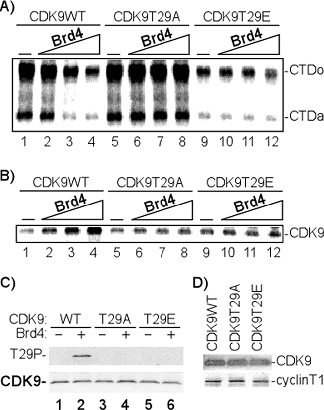
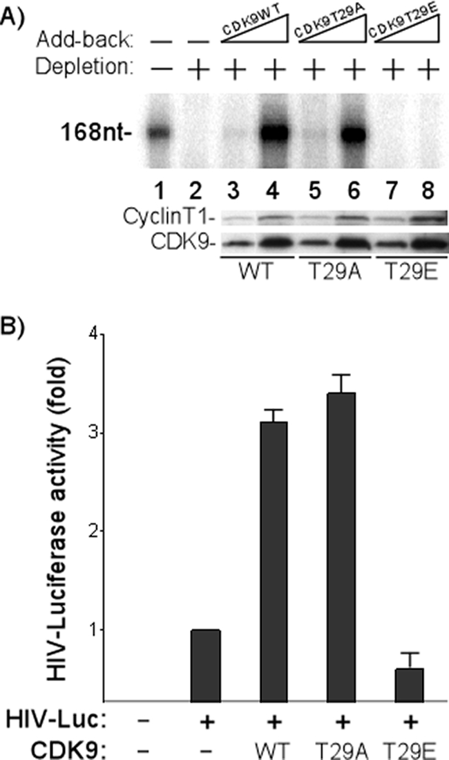
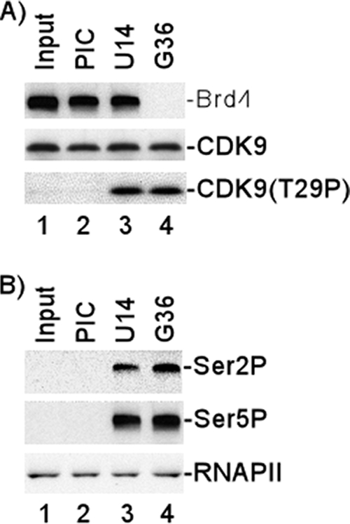
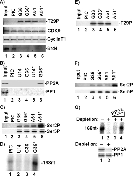
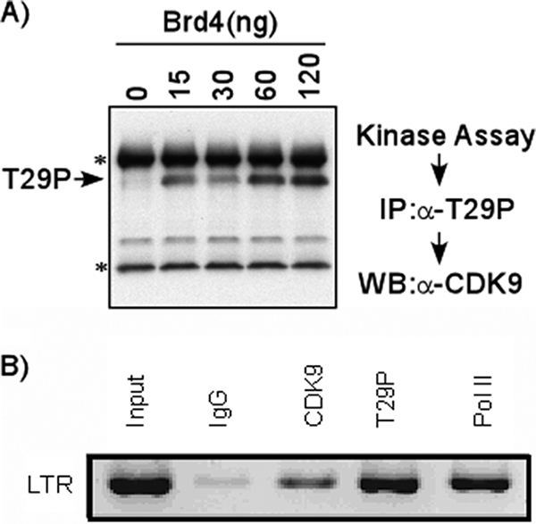
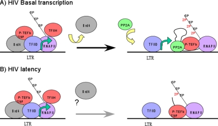
Positive transcription elongation factor b (P-TEFb), composed of cyclin-dependent kinase 9 (CDK9) and cyclin T, is a global transcription factor for eukaryotic gene expression, as well as a key factor for human immunodeficiency virus (HIV) transcription elongation. P-TEFb phosphorylates the carboxyl-terminal domain (CTD) of the large subunit of RNA polymerase II (RNAP II), facilitating the transition from nonprocessive to processive transcription elongation. Recently, the bromodomain protein Brd4 has been shown to interact with the low-molecular-weight, active P-TEFb complex and recruit P-TEFb to the HIV type 1 long terminal repeat (LTR) promoter. However, the subsequent events through which Brd4 regulates CDK9 kinase activity and RNAP II-dependent transcription are not clearly understood. Here we provide evidence that Brd4 regulates P-TEFb kinase activity by inducing a negative pathway. Moreover, by analyzing stepwise initiation and elongation complexes, we demonstrate that P-TEFb activity is regulated in the transcription complex. Brd4 induces phosphorylation of CDK9 at threonine 29 (T29) in the HIV transcription initiation complex, inhibiting CDK9 kinase activity. P-TEFb inhibition is transient, as Brd4 is released from the transcription complex between positions +14 and +36. Removal of the phosphate group at T29 by an incoming phosphatase released P-TEFb activity, resulting in increased RNAP II CTD phosphorylation and transcription. Finally, we present chromatin immunoprecipitation studies showing that CDK9 with phosphorylated T29 is associated with the HIV promoter region in the integrated and transcriptionally silent HIV genome.


Figures

FIG. 1.
FIG. 1.
Brd4 regulates CDK9 kinase activity through phosphorylation of Thr 29. (A) Effect of Brd4 on CTD phosphorylation by P-TEFb (CDK9). In vitro kinase assays were performed by incubating 50 ng GST-CTD; 50 ng P-TEFb, composed of WT CDK9 or mutants (T29A or T29E); and cyclin T1, with [γ-32P]ATP in the absence (−) or presence of increasing concentrations of Brd4 (30, 60, and 120 ng). The 32P-labeled GST-CTD was precipitated with glutathione-Sepharose beads and fractionated by electrophoresis on an 8% SDS-polyacrylamide gel, followed by autoradiography. The hypophosphorylated (CTDa) and hyperphosphorylated (CTDo) forms of CTD are indicated. The results shown are representative of three independent experiments performed under similar conditions. (B) Effect of Brd4 on CDK9 autophosphorylation. In vitro kinase assays were performed by incubating 50 ng P-TEFb with [γ-32P]ATP in the absence (−) or presence of increasing concentrations of Brd4 (30, 60, and 120 ng). 32P-labeled CDK9 was immunoprecipitated with anti-CDK9 antibody and analyzed by electrophoresis on a 4-to-20% SDS-polyacrylamide gel, followed by autoradiography. The results shown are representative of three independent experiments performed under similar conditions. (C) CDK9 T29 phosphorylation in the presence of Brd4. In vitro kinase assays were performed by incubating 50 ng P-TEFb, composed of WT CDK9 or mutants (T29A or T29E) and cyclin T1, with ATP in the absence (−) or presence (+) of 120 ng Brd4. CDK9 T29 phosphorylation was analyzed by electrophoresis on a 4-to-20% SDS-polyacrylamide gel, followed by Western blotting with anti-CDK9 or anti-T29P antibody. (D) Proteins used in the kinase assays whose results are shown in panels A and B were analyzed by Western blotting with anti-CDK9 or anti-cyclin T1, respectively.
FIG. 2.
FIG. 2.
CDK9 T29 phosphorylation inhibits transcription activity of P-TEFb in vitro and in vivo. (A) CDK9 T29 phosphorylation inhibits in vitro transcription. P-TEFb was depleted (+; −, not depleted) from HeLa nuclear extracts with anti-CDK9 antibody. Increasing concentrations of baculovirus-purified P-TEFb (50 ng and 150 ng) containing WT, T29A, or T29E CDK9 were then added back to the depleted extracts (−, no add-back). In vitro transcription reactions were set up by incubating the HIV template, reconstituted nuclear extracts, 50 μM ATP, 50 μM CTP, 50 μM GTP, 1.25 μM UTP, 20 μCi[α-32P]UTP, and 10 units of RNasin (Promega) in 1× IVT buffer. The radiolabeled transcripts (168 nucleotides [168nt]) (47) were fractionated by electrophoresis on a 6% denaturing polyacrylamide gel, followed by autoradiography. Western blot analyses of WT and mutant P-TEFb complexes added back to the depleted extracts are shown in the lower panels. (B) CDK9 T29 phosphorylation regulates P-TEFb transcription activity in vivo. The plasmids containing CDK9 WT or mutants were cotransfected into HeLa cells with a plasmid containing the HIV LTR-driven luciferase (HIV-Luc). The cells were cultured for 48 h and luciferase activity was assayed. Each result shown is the average of the results of four experiments with the standard error indicated. +, present; −, absent.
FIG. 3.
FIG. 3.
Association of Brd4 with HIV transcription complexes and CDK9 T29 phosphorylation during transcription. HIV-1 PICs were assembled by incubating biotinylated HIV-1 templates with HeLa nuclear extract and then purified with streptavidin-coated magnetic beads. The purified PICs were incubated with 50 μM ATP for 10 min and then washed extensively with 1× IVT buffer. The PICs were walked to position +U14 by incubation with 50 μM CTP, GTP, and UTP for 5 min at 30°C and then washed extensively with 1× IVT buffer. The TECs stalled at U14 were walked stepwise along the DNA by repeated incubation with different sets of three NTPs and then washed extensively with 1× IVT buffer to remove the unincorporated NTPs. (A) Protein compositions of PICs and TECs stalled at different stages were analyzed by fractionation by electrophoresis on 4-to-20% SDS-polyacrylamide gels, followed by Western blot analyses with antibody against Brd4, CDK9, or T29P CDK9. (B) Protein compositions of PICs and TECs stalled at different stages were analyzed by fractionation by electrophoresis on 4% SDS-polyacrylamide gels, followed by Western blot analyses with antibody against Ser 2P RNAP II CTD or Ser 5P RNAP II CTD. Western blot analysis with specific antibody (N-20) against the amino terminus of the largest subunit of RNAP II shown in bottom panel demonstrated equal amounts of RNAP II in the PICs and TECs.
FIG. 4.
FIG. 4.
Analysis of T29P in HIV transcription initiation complex and TECs. (A) State of CDK9 T29 phosphorylation. The purified PICs were walked to position +U14 by incubation with 50 μM CTP, GTP, and UTP for 5 min at 30°C and then washed extensively with 1× IVT buffer. The TECs stalled at U14 were walked stepwise along the DNA by repeated incubation with different sets of three NTPs and then washed extensively with 1× IVT buffer to remove the unincorporated NTPs. TECs stalled at G36 were reincubated without (G36) or with (G36*) RNAP II-depleted extract and then assayed directly or elongated stepwise to position +51 (A51, A51*). The protein compositions of PICs and TECs that were stalled at different stages were analyzed by Western blotting with anti-T29P, anti-CDK9, anti-cyclin T1, or anti-Brd4. (B) PP2A was recruited into TECs. The purified PICs were walked to position +U14 by incubation with 50 μM CTP, GTP, and UTP for 5 min at 30°C and then washed extensively with 1× IVT buffer. The TECs stalled at U14 were walked stepwise along the DNA by repeated incubation with different sets of three NTPs and then washed extensively with 1× IVT buffer to remove the unincorporated NTPs. TECs stalled at U14 or G36 were reincubated with RNAP II-depleted extracts (U14* or G36*, respectively) and then washed extensively with 1× IVT buffer. The protein compositions of PICs and TECs that were stalled at different stages were analyzed by Western blotting with anti-PP2A or anti-PP1. (C) Analysis of RNAP II CTD phosphorylation. The protein compositions of PICs and TECs that were stalled at different stages were analyzed by Western blotting with antibody against Ser 2P RNAP II CTD or Ser 5P RNAP II CTD. (D) Recruitment of phosphatase correlates with HIV transcription in vitro. Runoff transcripts (168nt) from TECs that were stalled at different stages were fractionated by electrophoresis on a 6% denaturing polyacrylamide gel, followed by autoradiography. (E and F) Purified PP2A dephosphorylates T29P during transcription elongation. The purified PICs were walked to position +U14 by incubation with 50 μM CTP, GTP, and UTP for 5 min at 30°C and then washed extensively with 1× IVT buffer. The TECs stalled at U14 were walked stepwise along the DNA by repeated incubation with different sets of three NTPs and then washed extensively with 1× IVT buffer to remove the unincorporated NTPs. TECs stalled at G36 were reincubated without (G36) or with (G36*) purified PP2A and then elongated stepwise to position +51 (A51, A51*). The protein compositions of PICs and TECs that were stalled at different stages were analyzed by Western blotting with anti-T29P (E) or anti-Ser 2P RNAP II CTD or anti-Ser 5P RNAP II CTD (F). (G) PP2A was required for the basal transcription of HIV. PP2A was added back in increasing concentrations (25 ng and 50 ng) to PP2A-depleted extract (+; −, not depleted), and then in vitro transcription assays were performed by incubating HIV templates with the reconstituted extracts. The radiolabeled transcripts (168nt) were fractionated by electrophoresis on a 6% denaturing polyacrylamide gel and detected by autoradiography. Western blot analyses of the PP2A-depleted extract are shown in the lower panels.
FIG. 5.
FIG. 5.
T29P CDK9 associated with latent HIV promoter in vivo. (A) Specificity of T29P CDK9 antibody for IP. In vitro kinase assays were performed by incubating P-TEFb with ATP in the presence of different concentrations of Brd4. The phosphorylated CDK9 was then immunoprecipitated with T29P CDK9 antibody (rabbit) and fractionated by electrophoresis on a 4-to-20% SDS-polyacrylamide gel, followed by Western blot analysis with anti-CDK9 antibody (mouse). Bands indicated by asterisks represent heavy and light chains of the antibody used for IP. α, anti; WB, Western blot. (B) T29P CDK9 is associated with latent HIV promoter in vivo. ChIP assays were carried out with TZM-bl cells, which contain separate integrated copies of the luciferase and β-galactosidase genes under the control of the HIV-1 promoter. Cross-linked extracts were immunoprecipitated with 5 μg of control IgG, anti-CDK9, anti-T29P CDK9, or anti-RNAP II (Pol II) antibody. PCR analysis with primers specific for the HIV LTR region was performed on the precipitated DNA.
FIG. 6.
FIG. 6.
Model for Brd4 regulation of P-TEFb activity. (A and B) Schematic diagrams of basal transcription of HIV and HIV latency are shown. Brd4 recruits P-TEFb to the HIV promoter. However, as the complex proceeds from initiation to elongation, Brd4 induces autophosphorylation of P-TEFb at threonine 29 (T29P), inhibiting P-TEFb CTD phosphorylation. In the case of basal transcription, Brd4 exits the complex, allowing the phosphatase PP2A to enter the complex, dephosphorylate P-TEFb, and relieve the inhibition of its kinase activity. This relieves the block to elongation and allows basal gene expression (A). In the case of HIV latency, P-TEFb remains phosphorylated at T29 and elongation cannot proceed (B).

References

    1. Ammosova, T., K. Washington, Z. Debebe, J. Brady, and S. Nekhai. 2005. Dephosphorylation of CDK9 by protein phosphatase 2A and protein phosphatase-1 in Tat-activated HIV-1 transcription. Retrovirology 247. - PMC - PubMed
    1. Bisgrove, D. A., T. Mahmoudi, P. Henklein, and E. Verdin. 2007. Conserved P-TEFb-interacting domain of BRD4 inhibits HIV transcription. Proc. Natl. Acad. Sci. USA. 10413690-13695. - PMC - PubMed
    1. Buratowski, S. 2003. The CTD code. Nat. Struct. Biol. 10679-680. - PubMed
    1. Dey, A., F. Chitsaz, A. Abbasi, T. Misteli, and K. Ozato. 2003. The double bromodomain protein Brd4 binds to acetylated chromatin during interphase and mitosis. Proc. Natl. Acad. Sci. USA. 1008758-8763. - PMC - PubMed
    1. Dey, A., J. Ellenberg, A. Farina, A. E. Coleman, T. Maruyama, S. Sciortino, J. Lippincott-Schwartz, and K. Ozato. 2000. A bromodomain protein, MCAP, associates with mitotic chromosomes and affects G2-to-M transition. Mol. Cell. Biol. 206537-6549. - PMC - PubMed

LinkOut - more resources