. 2009 Jan;83(2):498-511.
doi: 10.1128/JVI.01376-08. Epub 2008 Oct 29.

The addition of tumor necrosis factor plus beta interferon induces a novel synergistic antiviral state against poxviruses in primary human fibroblasts

Affiliations

The addition of tumor necrosis factor plus beta interferon induces a novel synergistic antiviral state against poxviruses in primary human fibroblasts

Eric Bartee et al. J Virol. 2009 Jan.

Abstract

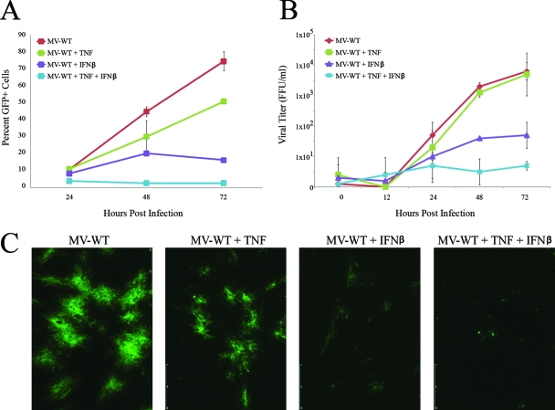
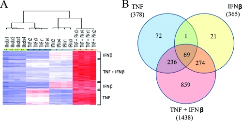
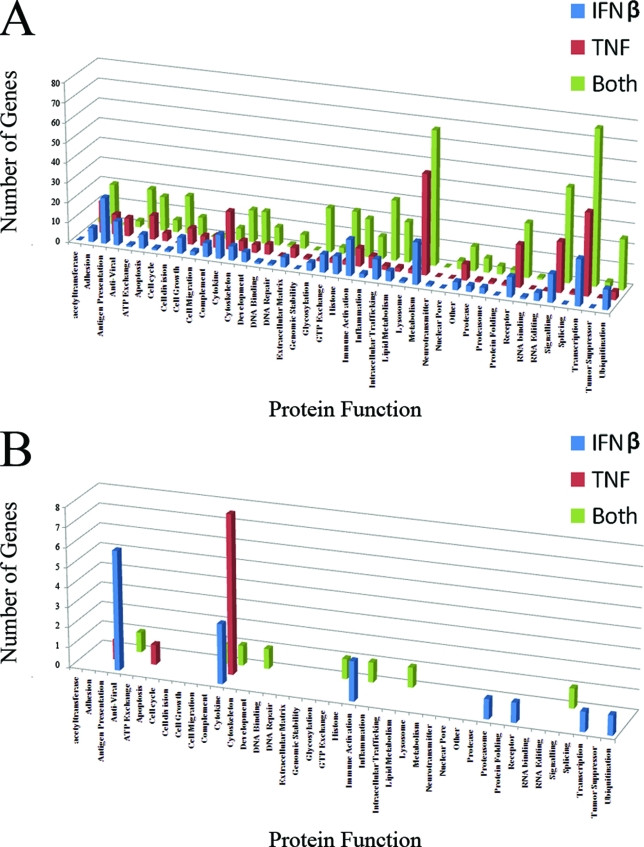
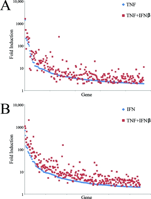
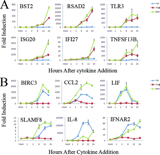
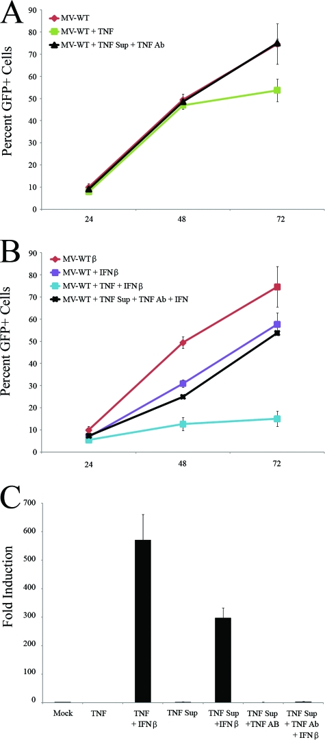
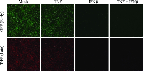
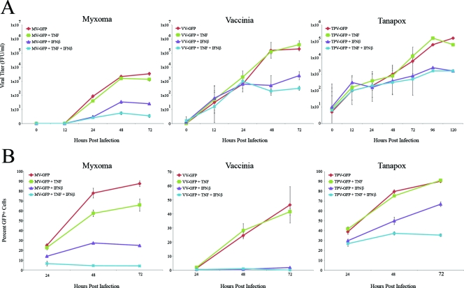
Tumor necrosis factor (TNF) and members of the interferon (IFN) family have been shown to independently inhibit the replication of a variety of viruses. In addition, previous reports have shown that treatment with various combinations of these antiviral cytokines induces a synergistic antiviral state that can be significantly more potent than addition of any of these cytokines alone. The mechanism of this cytokine synergy and its effects on global gene expression, however, are not well characterized. Here, we use DNA microarray analysis to demonstrate that treatment of uninfected primary human fibroblasts with TNF plus IFN-beta induces a distinct synergistic state characterized by significant perturbations of several hundred genes which are coinduced by the individual cytokines alone, as well as the induction of more than 850 novel host cell genes. This synergy is mediated directly by the two ligands, not by intermediate secreted factors, and is necessary and sufficient to completely block the productive replication and spread of myxoma virus in human fibroblasts. In contrast, the replication of two other poxviruses, vaccinia virus and tanapox virus, are only partially inhibited in these cells by the synergistic antiviral state, whereas the spread of both of these viruses to neighboring cells was efficiently blocked. Taken together, our data indicate that the combination of TNF and IFN-beta induces a novel synergistic antiviral state that is highly distinct from that induced by either cytokine alone.


Figures

FIG. 1.
FIG. 1.
TNF and IFN-β synergistically inhibit MV in primary human fibroblasts. Primary human GM02504 fibroblasts were infected with MV-GFP at an MOI of 0.1. After 1 h of viral adsorption, cells were treated with TNF, IFN-β, or the combination of TNF plus IFN-β. To determine the effect of these treatments on viral spread, cells were harvested via trypsin treatment at 24, 48, and 72 h postinfection. The percentage of GFP+ live cells in each sample was then determined by using flow cytometry. (A) Graph depicting a representative experiment run in triplicate. (B) Graph depicting the average of three independent experiments. (C) Prior to each analysis of each experiment, focus sizes were observed by florescence microscopy. To determine the effect of each treatment on virus titer, cells were harvested at the given time points via trypsinization. Cells were then lysed via repeated freeze-thawing, and virus titers were determined as outlined in Materials and Methods.
FIG. 2.
FIG. 2.
Treatment with TNF plus IFN-β causes upregulation of a large set of human genes not induced by TNF or IFN-β alone. An f-test was preformed to identify probe sets perturbed significantly (P < 0.001) among the four treatment groups. (A) The heat map displays the probe sets from this analysis, which also displayed a level of induction of >2-fold compared to mock treatment groups. The general grouping of these genes into sets that are induced by TNF, IFN-β, or TNF plus IFN-β is shown on the right of the heat map. Inspection of the dendrogram obtained after hierarchical clustering showed that for all replicates of each treatment class clustered tightly together. (B) Venn diagram analysis to illustrate the number of genes that overlapped between multiple treatment classes. Note that the Venn diagram displays genes that were significantly perturbed from the mock-treated sample (P < 0.001).
FIG. 3.
FIG. 3.
Functional categories of upregulated human genes. Each human gene that was scored as upregulated in the DNA microarray analysis was grouped into a functional category based on its annotation in the Swiss-Prot database. Graphs depict the total number of genes in each category (A), as well as the number of genes in each category which were significant at P < 0.001 and were upregulated by >50-fold (B).
FIG. 4.
FIG. 4.
Treatment with TNF plus IFN-β results in “synergy by cooperative action.” (A) Graph depicting the 236 human genes upregulated by TNF alone or TNF plus IFN-β; (B) graph depicting the 274 human genes upregulated by either IFN-β alone or TNF plus IFN-β. Each gene is shown twice on each graph. The fold change induced by the single cytokine alone is shown in blue, while the fold change induced by the combination of both cytokines is shown in red.
FIG. 5.
FIG. 5.
The combination of TNF plus IFN-β alters the kinetics of human gene induction. Primary human GM02504 fibroblasts were treated with TNF, IFN-β, or TNF plus IFN-β. At the listed time points, RNA was extracted from each sample and used to synthesize cDNA. The expression of each gene at each time point was assayed with gene-specific primers using Sybr green-based real-time PCR as detailed in Materials and Methods. The graphs in panels A and B show the average of two independent experiments each done in triplicate.
FIG. 6.
FIG. 6.
Synergy between TNF and IFN-β is not mediated by a secreted intermediate factor. To generate TNF-treated supernatants, GM02504 primary human fibroblasts were treated with 1 ng of TNF/ml for 24 h. TNF was then removed from the resulting supernatant by using a depleting anti-huTNF antibody. To determine whether this TNF-depleted supernatant had any anti-MV properties, fresh GM02504 cells were infected with MV-GFP at an MOI of 0.1 and then either mock treated, treated with 1 ng of TNF/ml, or treated with the TNF supernatant. (A) At 24, 48, and 72 h postinfection, cells were harvested, and the ability of MV-GFP to spread cell to cell was assayed by flow cytometry. To determine whether secreted factor(s) in the TNF-treated supernatant was able to synergize with IFN-β, fresh GM02504 cells were infected with MV at an MOI of 0.1 and then either mock treated or treated with IFN-β, IFN-β + 1 ng of TNF/ml, or IFN-β + TNF supernatant. (B) At 24, 48, and 72 h, the ability of MV-GFP to spread through these cultures was assayed by using flow cytometry. To further analyze whether the TNF supernatants could induce synergy with IFN-β, the ability of these supernatants to induce “synergy by cooperative induction” was tested by using real-time PCR. Fresh GM02504 cells were treated as indicated. At 24 h after treatment, RNA was harvested from cells and used to make cDNA. Levels of gene expression were assayed with gene specific primers using Sybr green-based real-time PCR as detailed in Materials and Methods. (C) Graph showing the induction of CXCL10.
FIG. 7.
FIG. 7.
The combination of TNF plus IFN-β blocks MV replication prior to, or at the level of, early gene expression. GM02504 primary human fibroblasts were treated with TNF, IFN-β, or TNF plus IFN-β. At 24 h after the addition of these cytokines, cells were infected with a recombinant MV that expresses both GFP, under an sE/L promoter, and TrFP, under a viral P11 late promoter. At 24 h after infection, the levels of both GFP and TrFP were measured via florescence microscopy.
FIG. 8.
FIG. 8.
The combination of TNF plus IFN-β uniquely affects multiple poxviruses. Primary human GM02504 fibroblasts were infected with MV-GFP, VV-GFP, or TPV-GFP at an MOI of 0.1. After 1 h of viral adsorption, cells were treated with TNF, IFN-β, or the combination of TNF plus IFN-β. To determine the effect of each treatment on the progeny virus titer, cells were harvested at the given time points via trypsinization. Cells were then lysed via repeated freeze-thawing, and virus titers were determined as outlined in Materials and Methods. (A) Graph depicting the average of three independent experiments. To determine the effect of these treatments on viral spread, cells were harvested via trypsinization at 24, 48, and 72 h postinfection. The percentage of GFP+ live cells in each sample was then determined by using flow cytometry. (B) Graph depicting a representative experiment run in triplicate.

References

    1. Adam, R., D. Russing, O. Adams, A. Ailyati, K. Sik Kim, H. Schroten, and W. Daubener. 2005. Role of human brain microvascular endothelial cells during central nervous system infection: significance of indoleamine 2,3-dioxygenase in antimicrobial defense and immunoregulation. Thromb. Haemost. 94341-346. - PubMed
    1. Anderson, S. L., J. M. Carton, J. Lou, L. Xing, and B. Y. Rubin. 1999. Interferon-induced guanylate binding protein-1 (GBP-1) mediates an antiviral effect against vesicular stomatitis virus and encephalomyocarditis virus. Virology 2568-14. - PubMed
    1. Bartee, E., M. R. Mohamed, and G. McFadden. 2008. Tumor necrosis factor and interferon: cytokines in harmony. Curr. Opin. Microbiol. 11378-383. - PMC - PubMed
    1. Carter, C. C., V. Y. Gorbacheva, and D. J. Vestal. 2005. Inhibition of VSV and EMCV replication by the interferon-induced GTPase, mGBP-2: differential requirement for wild-type GTP binding domain. Arch. Virol. 1501213-1220. - PubMed
    1. Chiu, J. J., P. L. Lee, S. F. Chang, L. J. Chen, C. I. Lee, K. M. Lin, S. Usami, and S. Chien. 2005. Shear stress regulates gene expression in vascular endothelial cells in response to tumor necrosis factor-alpha: a study of the transcription profile with complementary DNA microarray. J. Biomed. Sci. 12481-502. - PubMed

Publication types

MeSH terms

LinkOut - more resources