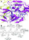
Mechanism of NS2B-mediated activation of NS3pro in dengue virus: molecular dynamics simulations and bioassays
- PMID: 18971276
- PMCID: PMC2612360
- DOI: 10.1128/JVI.01325-08
Mechanism of NS2B-mediated activation of NS3pro in dengue virus: molecular dynamics simulations and bioassays
Abstract
The NS2B cofactor is critical for proteolytic activation of the flavivirus NS3 protease. To elucidate the mechanism involved in NS2B-mediated activation of NS3 protease, molecular dynamic simulation, principal component analysis, molecular docking, mutagenesis, and bioassay studies were carried out on both the dengue virus NS3pro and NS2B-NS3pro systems. The results revealed that the NS2B-NS3pro complex is more rigid than NS3pro alone due to its robust hydrogen bond and hydrophobic interaction networks within the complex. These potent networks lead to remodeling of the secondary and tertiary structures of the protease that facilitates cleavage sequence recognition and binding of substrates. The cofactor is also essential for proper domain motion that contributes to substrate binding. Hence, the NS2B cofactor plays a dual role in enzyme activation by facilitating the refolding of the NS3pro domain as well as being directly involved in substrate binding/interactions. Kinetic analyses indicated for the first time that Glu92 and Asp50 in NS2B and Gln27, Gln35, and Arg54 in NS3pro may provide secondary interaction points for substrate binding. These new insights on the mechanistic contributions of the NS2B cofactor to NS3 activation may be utilized to refine current computer-based search strategies to raise the quality of candidate molecules identified as potent inhibitors against flaviviruses.
Figures


Similar articles
-
Structural basis for the activation of flaviviral NS3 proteases from dengue and West Nile virus.Nat Struct Mol Biol. 2006 Apr;13(4):372-3. doi: 10.1038/nsmb1073. Epub 2006 Mar 12. Nat Struct Mol Biol. 2006. PMID: 16532006
-
The active essential CFNS3d protein complex.FEBS J. 2006 Aug;273(16):3650-62. doi: 10.1111/j.1742-4658.2006.05369.x. FEBS J. 2006. PMID: 16911516
-
Homology modeling and molecular dynamics simulations of Dengue virus NS2B/NS3 protease: insight into molecular interaction.J Mol Recognit. 2010 May-Jun;23(3):283-300. doi: 10.1002/jmr.977. J Mol Recognit. 2010. PMID: 19693793
-
Identification of residues in the dengue virus type 2 NS2B cofactor that are critical for NS3 protease activation.J Virol. 2004 Dec;78(24):13708-16. doi: 10.1128/JVI.78.24.13708-13716.2004. J Virol. 2004. PMID: 15564480 Free PMC article.
-
Towards the design of antiviral inhibitors against flaviviruses: the case for the multifunctional NS3 protein from Dengue virus as a target.Antiviral Res. 2008 Nov;80(2):94-101. doi: 10.1016/j.antiviral.2008.07.001. Epub 2008 Jul 30. Antiviral Res. 2008. PMID: 18674567 Review.
Cited by
-
Mitochondrial Import of Dengue Virus NS3 Protease and Cleavage of GrpEL1, a Cochaperone of Mitochondrial Hsp70.J Virol. 2020 Aug 17;94(17):e01178-20. doi: 10.1128/JVI.01178-20. Print 2020 Aug 17. J Virol. 2020. PMID: 32581108 Free PMC article.
-
Host Factor SPCS1 Regulates the Replication of Japanese Encephalitis Virus through Interactions with Transmembrane Domains of NS2B.J Virol. 2018 May 29;92(12):e00197-18. doi: 10.1128/JVI.00197-18. Print 2018 Jun 15. J Virol. 2018. PMID: 29593046 Free PMC article.
-
NMR and MD Studies Reveal That the Isolated Dengue NS3 Protease Is an Intrinsically Disordered Chymotrypsin Fold Which Absolutely Requests NS2B for Correct Folding and Functional Dynamics.PLoS One. 2015 Aug 10;10(8):e0134823. doi: 10.1371/journal.pone.0134823. eCollection 2015. PLoS One. 2015. PMID: 26258523 Free PMC article.
-
Transmembrane Domains of NS2B Contribute to both Viral RNA Replication and Particle Formation in Japanese Encephalitis Virus.J Virol. 2016 May 27;90(12):5735-5749. doi: 10.1128/JVI.00340-16. Print 2016 Jun 15. J Virol. 2016. PMID: 27053551 Free PMC article.
-
Potential Role of Flavivirus NS2B-NS3 Proteases in Viral Pathogenesis and Anti-flavivirus Drug Discovery Employing Animal Cells and Models: A Review.Viruses. 2021 Dec 28;14(1):44. doi: 10.3390/v14010044. Viruses. 2021. PMID: 35062249 Free PMC article. Review.
References
-
- Amadei, A., A. B. Linssen, and H. J. Berendsen. 1993. Essential dynamics of proteins. Proteins 17412-425. - PubMed
-
- Arias, C. F., F. Preugschat, and J. H. Strauss. 1993. Dengue 2 virus NS2B and NS3 form a stable complex that can cleave NS3 within the helicase domain. Virology 193888-899. - PubMed
-
- Berendsen, H. J. C., J. P. M. Postma, W. F. van Gunsteren, A. DiNola, and J. R. Haak. 1984. Molecular dynamics with coupling to an external bath. J. Chem. Phys. 813684-3690.
-
- Berendsen, H. J. C., D. van der Spoel, and R. van Drunen. 1995. GROMACS: a message-passing parallel molecular dynamics implementation. Comp. Phys. Commun. 9143-56.
-
- Bessaud, M., B. A. Pastorino, C. N. Peyrefitte, D. Rolland, M. Grandadam, and H. J. Tolou. 2006. Functional characterization of the NS2B/NS3 protease complex from seven viruses belonging to different groups inside the genus Flavivirus. Virus Res. 12079-90. - PubMed
Publication types
MeSH terms
Substances
LinkOut - more resources
Full Text Sources

