. 2009 Jan;83(1):128-39.
doi: 10.1128/JVI.01954-08. Epub 2008 Oct 29.

Identification of sequences in herpes simplex virus type 1 ICP22 that influence RNA polymerase II modification and viral late gene expression

Affiliations

Identification of sequences in herpes simplex virus type 1 ICP22 that influence RNA polymerase II modification and viral late gene expression

Thomas W Bastian et al. J Virol. 2009 Jan.

Abstract

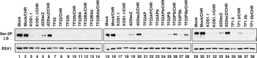
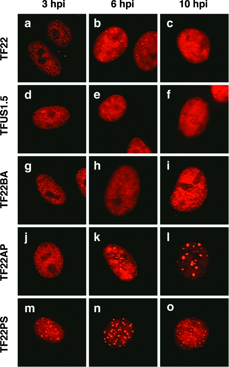
Previous studies have shown that the herpes simplex virus type 1 (HSV-1) immediate-early protein ICP22 alters the phosphorylation of the host cell RNA polymerase II (Pol II) during viral infection. In this study, we have engineered several ICP22 plasmid and virus mutants in order to map the ICP22 sequences that are involved in this function. We identify a region in the C-terminal half of ICP22 (residues 240 to 340) that is critical for Pol II modification and further show that the N-terminal half of the protein (residues 1 to 239) is not required. However, immunofluorescence analysis indicates that the N-terminal half of ICP22 is needed for its localization to nuclear body structures. These results demonstrate that ICP22's effects on Pol II do not require that it accumulate in nuclear bodies. As ICP22 is known to enhance viral late gene expression during infection of certain cultured cells, including human embryonic lung (HEL) cells, we used our engineered viral mutants to map this function of ICP22. It was found that mutations in both the N- and C-terminal halves of ICP22 result in similar defects in viral late gene expression and growth in HEL cells, despite having distinctly different effects on Pol II. Thus, our results genetically uncouple ICP22's effects on Pol II from its effects on viral late gene expression. This suggests that these two functions of ICP22 may be due to distinct activities of the protein.


Figures

FIG. 1.
FIG. 1.
Expression of mutant ICP22 polypeptides in transfected cells. (A) Diagram of modified ICP22 polypeptides. The bar at the top represents the 420-residue ICP22 polypeptide. The positions of known nuclear localization signals (NLSs) (residues 16 to 31 and 118 to 131) (54) are shown, as is a region conserved among alphaherpesviral ICP22 homologs (residues 161 to 292). The solid black bars below show the ICP22 protein sequences present in the various mutants, with the white bar representing the N-terminal FLAG epitope (F). Dotted lines indicate deletions. The restriction enzymes used to generate the deletions are indicated (BmgBI [B], AleI [A], PmlI [P], and SacII [S]). (B) Immunoblot analysis of transiently expressed proteins. Vero cells were transfected with the indicated plasmids, and protein extracts were prepared after 24 h. The proteins were analyzed by immunoblotting using antisera specific for ICP22 or FLAG. α-ICP22, anti-ICP22 antibody. (C) Localization of mutant polypeptides. Vero cells were transfected with the various plasmids as indicated and processed for immunofluorescence 1 day posttransfection using FLAG-specific antisera. Plasmids pcDNA22 (22), pcDNAUS1.5 (US1.5), pcDNA22-BA (BA), pcDNA22-AP (AP), and pcDNA22-PS (PS) were used. Representative cells are shown. (D) ICP22-triggered loss of Ser2-P Pol II. Vero cells were transfected with pCMVβgal-c (a) or the ICP22 expression plasmids as indicated (b to f), and processed for immunofluorescence 24 h later. Cells were doubly stained for β-galactosidase (β-gal) (green signal) and Ser-2P Pol II (red signal) (a), or ICP22 (green signal) and Ser-2P Pol II (red signal) (b to f). The panels show the merged green and red images.
FIG. 2.
FIG. 2.
Characterization of viral ICP22 mutants. (A) Expected genome structures of viral mutants. At the top is shown a diagram of the HSV-1 genome as well as a blow-up of the ICP22 gene contained on the BamHI N restriction fragment. The spliced US1 transcript is indicated. The white and black bars denote the HSV-1 genome repeats RL and RS, respectively. The sizes (in kilobases) of expected BamHI fragments in the region of the ICP22 gene are shown; the parentheses denote deletions. B, BamHI restriction sites. (B) Southern blot analysis. Total DNA was purified from infected Vero cells, digested with BamHI, and analyzed by Southern blotting using 32P-labeled pBamN as a probe. The positions of DNA size standards are shown to the left of the blot. The asterisk denotes the 1.9-kb BamHI fragment arising from the opposite US/RS junction. (C) Immunoblotting analysis. Vero cells were mock infected or infected with the indicated viruses at an MOI of 10. Protein extracts were prepared at 6 hpi, and immunoblotting was carried using ICP22- or FLAG-specific antisera. The migration positions of protein molecular mass markers (in kilodaltons) are shown to the left of each image. α-ICP22, anti-ICP22 antibody.
FIG. 3.
FIG. 3.
Growth of ICP22 mutants in Vero and HEL cells. Confluent monolayers of Vero (A) or HEL (B) cells were infected in triplicate with the viruses indicated at an MOI of 10 and incubated for 24 h. Virus yield in the infected cells was determined by plaque assay of the cell lysates on Vero cells. Bars denote the mean virus yield; error bars represent the standard error of three triplicate infections.
FIG. 4.
FIG. 4.
Induction of Pol III in ICP22 mutant-infected cells. Vero cells were mock infected or infected with the indicated viruses at an MOI of 10. Protein extracts were prepared at 6 hpi and analyzed by immunoblotting using the Pol II LS-specific antibodies ARNA3 or 8WG16. The same samples were also probed for ICP27 and EEA1. The Pol II LS form IIa migrates at approximately 200 kDa; II0 migrates at approximately 240 kDa. The two blots show the results of separate experiments.
FIG. 5.
FIG. 5.
Loss of Ser-2P Pol II in ICP22 mutant-infected cells. Vero cells were mock infected or infected with the indicated viruses. In one set of infections (odd-number lanes), the cells were left untreated, and protein extracts were prepared at 7 hpi. In the other set of infections (labeled CHR; even-number lanes), cells were infected in the presence of 50 μg/ml CH for 5 h. The CH was then removed, and the cells were incubated for two more hours prior to harvesting. Protein extracts were analyzed by immunoblotting using the H5 antibody or an antibody specific for EEA1. The blots on the left and in the middle show the results of one experiment, whereas the blot on the right shows the results of a separate experiment.
FIG. 6.
FIG. 6.
Localization of ICP22 polypeptides during infection. Subconfluent monolayers of Vero cells were mock infected or infected with the mutants shown. At the times indicated, cells were fixed and processed for immunofluorescence using an anti-FLAG antibody.
FIG. 7.
FIG. 7.
Expression of DE/L proteins in ICP22 mutant-infected cells. HEL cells were mock infected or infected with the indicated viruses. At 8 hpi, protein extracts were prepared, and immunoblotting was carried out to detect the indicated viral DE/L proteins. Cellular protein EEA1 was analyzed as a loading control.
FIG. 8.
FIG. 8.
Conserved sequences in HSV-1 ICP22. (A) The bar represents the 420-residue ICP22 polypeptide. The region conserved among members of the subfamily Alphaherpesvirinae is indicated, as is as a C-terminal sequence conserved only in members of the genus Simplexvirus. The sequence deleted in the PS mutant is shown at the top. (B) Alignment of C-terminal sequence conserved in simplexviruses. The C-terminal sequences of ICP22 and four homologs from other simplexviruses (cercopithecine virus 2 [CeHV2] [simian agent 8], cercopithecine virus 16 [CeHV-16] [herpesvirus papio 2], cercopithecine virus 1 [CeHV-1] [monkey B virus], and herpes simplex virus type 2 [HSV-2]) were aligned using Clustalw (http://www.ebi.ac.uk/). Single and double dots indicate weakly and strongly conserved residues, respectively; asterisks indicate identical residues. Gaps introduced to maximize alignment are indicated by dashes.

Similar articles

Cited by

References

    1. Advani, S. J., R. Brandimarti, R. R. Weichselbaum, and B. Roizman. 2000. The disappearance of cyclins A and B and the increase in activity of the G2/M-phase cellular kinase cdc2 in herpes simplex virus 1-infected cells require expression of the α22/US1.5 and UL13 viral genes. J. Virol. 748-15. - PMC - PubMed
    1. Advani, S. J., R. R. Weichselbaum, and B. Roizman. 2003. Herpes simplex virus 1 activates cdc2 to recruit topoisomerase IIalpha for post-DNA synthesis expression of late genes. Proc. Natl. Acad. Sci. USA 1004825-4830. - PMC - PubMed
    1. Advani, S. J., R. R. Weichselbaum, and B. Roizman. 2000. The role of cdc2 in the expression of herpes simplex virus genes. Proc. Natl. Acad. Sci. USA 9710996-11001. - PMC - PubMed
    1. Cai, W. Z., S. Person, C. DebRoy, and B. H. Gu. 1988. Functional regions and structural features of the gB glycoprotein of herpes simplex virus type 1. An analysis of linker insertion mutants. J. Mol. Biol. 201575-588. - PubMed
    1. Carter, K. L., and B. Roizman. 1996. The promoter and transcriptional unit of a novel herpes simplex virus 1 α gene are contained in, and encode a protein in frame with, the open reading frame of the α22 gene. J. Virol. 70172-178. - PMC - PubMed

Publication types