. 2009 Jan;83(1):150-8.
doi: 10.1128/JVI.01652-08. Epub 2008 Oct 29.

Follicular dendritic cells and human immunodeficiency virus type 1 transcription in CD4+ T cells

Affiliations

Follicular dendritic cells and human immunodeficiency virus type 1 transcription in CD4+ T cells

Tyler C Thacker et al. J Virol. 2009 Jan.

Abstract

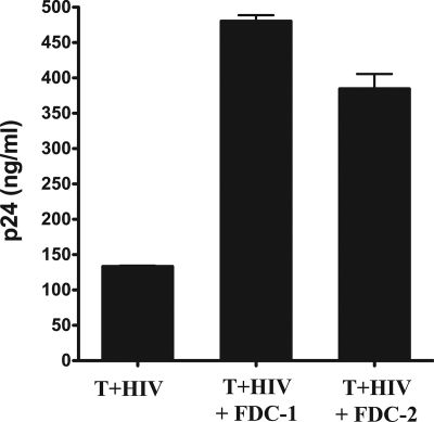
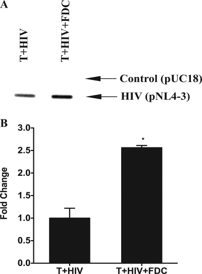
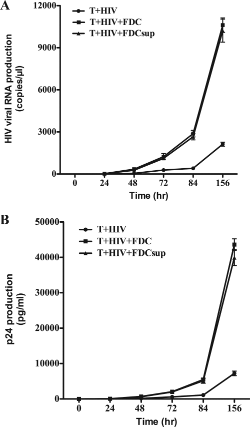
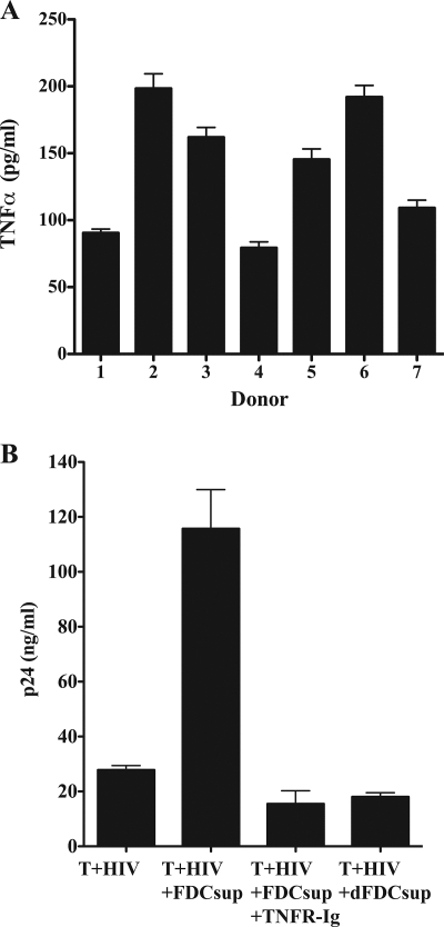
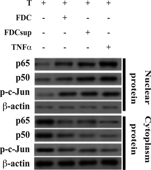
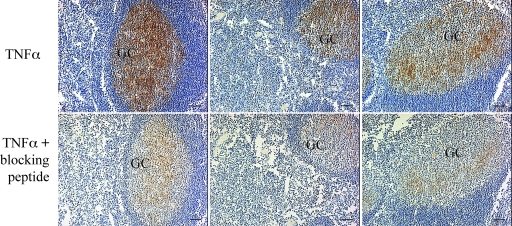
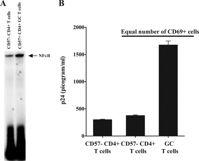
HIV replication occurs throughout the natural course of infection in secondary lymphoid tissues and in particular within the germinal centers (GCs), where follicular dendritic cells (FDCs) are adjacent to CD4(+) T cells. Because FDCs provide signaling that increases lymphocyte activation, we postulated that FDCs could increase human immunodeficiency virus (HIV) replication. We cultured HIV-infected CD4(+) T cells alone or with FDCs and measured subsequent virus expression using HIV-p24 production and reverse transcription-PCR analyses. When cultured with FDCs, infected CD4(+) T cells produced almost fourfold more HIV than when cultured alone, and the rate of virus transcription was doubled. Both FDCs and their supernatant increased HIV transcription and resulted in nuclear translocation of NF-kappaB and phosphorylated c-Jun in infected cells. FDCs produced soluble tumor necrosis factor alpha (TNF-alpha) ex vivo, and the addition of a blocking soluble TNF receptor ablated FDC-mediated HIV transcription. Furthermore, TNF-alpha was found highly expressed within GCs, and ex vivo GC CD4(+) T cells supported greater levels of HIV-1 replication than other CD4(+) T cells. These data indicated that FDCs increase HIV transcription and production by a soluble TNF-alpha-mediated mechanism. This FDC-mediated effect may account, at least in part, for the presence of persistent HIV replication in GCs. Therefore, in addition to providing an important reservoir of infectious virus, FDCs increase HIV production, contributing to a tissue microenvironment that is highly conducive to HIV transmission and expression.


Figures

FIG. 1.
FIG. 1.
FDCs cocultured with HIV-infected CD4+ T cells increased virus p24 production. In vitro-infected CD4+ T cells (T+HIV) were cultured for 6 days with or without FDCs (10 T cells per FDC). The tissue culture supernatant was then harvested and virus particle production measured by p24 ELISA. FDC-1 and FDC-2 refer to FDCs obtained from two separate, unrelated donors. All assays were performed in duplicate, and the error bars represent the SEM. Statistical significance (* signifies P values of <0.05) was determined by Student's t test. These data are representative of three independent experiments using different sources of primary CD4+ T cells and FDCs.
FIG. 2.
FIG. 2.
FDCs increased the rate of HIV transcription in HIV-infected T cells. (A) In vitro-infected CD4+ T cells were cultured with FDCs for 18 h. The nuclei were then isolated and subjected to nuclear run-on transcription analysis. A plasmid containing the full-length HIV genome (pNL4-3) was used to capture HIV-specific RNA while the parental plasmid without the HIV genome was used as a control for nonspecific binding. The Southern blot presented is representative of two independent experiments. (B) The density of the Southern blot was determined using ImageJ software. The differences between the control and pNL4-3 densities are plotted and represent the means for two independent experiments. The error bars represent the SEM. Statistical significance (* signifies P values of <0.05) was determined by Student's t test.
FIG. 3.
FIG. 3.
FDC augmentation of virus transcription was mediated by a soluble factor. In vitro-infected CD4+ T cells were cultured with FDCs or supernatant (FDCsup; 10%, vol/vol) obtained from 6-day cultures of FACS-sorted FDCs. (A) Viral RNA was harvested from the culture supernatants at the times indicated and examined using quantitative RT-PCR. (B) The supernatant fluid from the same cultures was harvested and examined for HIV p24 production at the times indicated. Error bars represent the standard deviations for replicate cultures. These data are representative of three independent experiments using different sources of primary CD4+ T cells and FDCs.
FIG. 4.
FIG. 4.
FDCs produce TNF-α that increases HIV replication. (A) FACS-sorted FDCs (1 × 10⁵/ml) from seven unrelated donors were cultured for 6 days, after which the culture supernatant fluid was examined for TNF-α production using ELISA. Data are expressed in picograms TNF-α per milliliter of culture fluid and represent the means ± standard deviations for triplicate wells. (B) In vitro-infected CD4+ T cells were cultured with supernatant (FDCsup) obtained from cultured FDCs with or without TNFR-Ig (40 μg/ml). As a negative control, the same cells were cultured with supernatant from secondary lymphoid tissue cells that had been specifically depleted of FDCs (dFDCsup). After 6 days of culture, the supernatants were assayed for virus production by p24 ELISA. The data are representative of two independent experiments using different donors of FDCs and T cells and represent the means for replicate cultures. The error bars represent the SEM.
FIG. 5.
FIG. 5.
FDCs induce activated NF-κB in in vitro-infected CD4+ T cells. FDCs (1 × 10⁵ cells/ml), supernatant fluid from FDC cultures (FDCsup; 10%, vol/vol), or recombinant TNF-α (166 picograms/ml) was cultured with in vitro-infected CD4+ T cells (1 × 10⁶ cells/ml) for 24 h, after which the cells were examined for cytoplasmic and nuclear NF-κB p50, p65, and phosphorylated c-Jun (p-c-Jun) by Western blotting as described in Materials and methods. β-Actin was also immunoblotted and served as a loading control. These data are representative of two independent experiments using different sources of primary CD4+ T cells and FDCs.
FIG. 6.
FIG. 6.
TNF-α is present in GCs. Tonsillar sections from three unrelated individuals were examined for the presence of TNF-α as described in Materials and Methods. Original magnification, ×100. The scale bar represents 50 μm.
FIG. 7.
FIG. 7.
Ex vivo GC CD4+ T cells are like CD4+ T cells cultured with FDCs in vitro. GC CD4+ T cells were isolated from the tonsils of healthy, uninfected donors as described in Materials and Methods. (A) GC T cells (CD4+ CD57+) express more activated NF-κB than non-GC CD4+ T cells obtained from the same tissue. Immediately after isolation, the nuclei were obtained from equal numbers of GC T cells (4 × 10⁶) and non-GC T cells and prepared and analyzed by an EMSA for NF-κB as described in Materials and Methods. (B) GC T cells infected in vitro produce more virus than other CD4+ T cells from the same tissue. Equal numbers (5 × 10⁵) of GC T cells and other CD4+ T cells from the same tissue were infected with HIV-1 92US714 (R5-tropic; 0.025 cpm reverse transcriptase activity) as described in Materials and Methods. After 6 days, the culture supernatant was harvested and the HIV produced assessed by p24 ELISA. These data are representative of three independent experiments. Because GC T cells express three- to fourfold more CXCR4 than other CD4+ T cells, we used an R5 primary isolate of virus for these studies to avoid potential differences due to virus binding and entry.

Similar articles

Cited by

References

    1. Armstrong, J. A. 1991. Ultrastructure and significance of the lymphoid tissue lesions in HIV infection, p. 69-82. In P. Racz, C. D. Dijkstra, and J. C. Gluckman (ed.), Accessory cells in HIV and other retroviral infections. Karger, Basel, Switzerland.
    1. Biberfeld, P., A. Porwit, G. Biberfield, M. Harper, A. Bodner, and R. Gallo. 1988. Lymphadenopathy in HIV (HTLV-III LAV) infected subjects: the role of virus and follicular dendritic cells. Cancer Detect. Prev. 12217-224. - PubMed
    1. Burton, G. F., B. F. Keele, J. D. Estes, T. C. Thacker, and S. Gartner. 2002. Follicular dendritic cell contributions to HIV pathogenesis. Semin. Immunol. 14275-284. - PubMed
    1. Burton, G. F., A. Masuda, S. L. Heath, B. A. Smith, J. G. Tew, and A. K. Szakal. 1997. Follicular dendritic cells (FDC) in retroviral infection: host/pathogen perspectives. Immunol. Rev. 156185-197. - PubMed
    1. Butch, A. W., G. H. Chung, J. W. Hoffmann, and M. H. Nahm. 1993. Cytokine expression by germinal center cells. J. Immunol. 15039-47. - PubMed

Publication types

MeSH terms