. 2009 Jan;83(1):314-28.
doi: 10.1128/JVI.01444-08. Epub 2008 Oct 29.

Alternatively initiated gene 50/RTA transcripts expressed during murine and human gammaherpesvirus reactivation from latency

Affiliations

Alternatively initiated gene 50/RTA transcripts expressed during murine and human gammaherpesvirus reactivation from latency

Kathleen S Gray et al. J Virol. 2009 Jan.

Abstract

In the process of characterizing the requirements for expression of the essential immediate-early transcriptional activator (RTA) encoded by gene 50 of murine gammaherpesvirus 68 (MHV68), a recombinant virus was generated in which the known gene 50 promoter was deleted (G50pKO). Surprisingly, the G50pKO mutant retained the ability to replicate in permissive murine fibroblasts, albeit with slower kinetics than wild-type MHV68. 5'-rapid amplification of cDNA ends analyses of RNA prepared from G50pKO-infected fibroblasts revealed a novel upstream transcription initiation site, which was also utilized during wild-type MHV68 infection of permissive cells. Furthermore, the region upstream of the distal gene 50/RTA transcription initiation site exhibited promoter activity in both permissive NIH 3T12 fibroblasts as well as in the murine macrophage cell line RAW 264.7. In addition, in RAW 264.7 cells the activity of the distal gene 50/RTA promoter was strongly upregulated (>20-fold) by treatment of the cells with lipopolysaccharide. Reverse transcriptase PCR analyses of RNA prepared from Kaposi's sarcoma-associated herpesvirus- and Epstein-Barr virus-infected B-cell lines, following induction of virus reactivation, also revealed the presence of gene 50/RTA transcripts initiating upstream of the known transcription initiation site. The latter argues that alternative initiation of gene 50/RTA transcription is a strategy conserved among murine and human gammaherpesviruses. Infection of mice with the MHV68 G50pKO demonstrated the ability of this mutant virus to establish latency in the spleen and peritoneal exudate cells (PECs). However, the G50pKO mutant was unable to reactivate from latently infected splenocytes and also exhibited a significant reactivation defect from latently infected PECs, arguing in favor of a model where the proximal gene 50/RTA promoter plays a critical role in virus reactivation from latency, particularly from B cells. Finally, analyses of viral genome methylation in the regions upstream of the proximal and distal gene 50/RTA transcription initiation sites revealed that the distal promoter is partially methylated in vivo and heavily methylated in MHV68 latently infected B-cell lines, suggesting that DNA methylation may serve to silence the activity of this promoter during virus latency.


Figures

FIG. 1.
FIG. 1.
Conservation of G50/BRLF1/Rta coding region among gammaherpesviruses. Schematic representation of the G50/BRLF1/Rta coding regions of MHV68, EBV, KSHV, and HVS. The nucleotide positions for G50 exons 1 and 2 are provided. The box drawn around exon 1 demonstrates the conserved juxtaposition of exon 1 relative to open reading frames encoded on the opposite strand.
FIG. 2.
FIG. 2.
Generation and confirmation of MHV68.G50pKO and MHV68.G50pKO-MR viruses. (A) Schematic depicting the organization of open reading frames in the G50 region. The upright arrow indicates the relative position of the characterized G50 promoter, and the hatched area indicates the region of the deletion in the MHV68.G50pKO virus. The indicated splicing of E1 to E2 gives rise to a full-length G50-encoding transcript. Wild-type sequence contains HindIII sites at bp 64898 and bp 57541; the MHV68.G50pKO BAC contains an additional HindIII site at 66549 (indicated by the asterisk). (B) HindIII digest of MHV68 wild-type, G50pKO, and G50pKO-MR virus BACs. The arrows indicate the fragments produced by the introduction of the additional HindIII site in panel A. (C) Southern blot hybridization using the PCR-generated probe indicated in panel A.
FIG. 3.
FIG. 3.
MHV68.G50pKO virus replicates in vitro. (A) Single-step growth analysis. NIH 3T12 cells were infected with wild-type or MHV68.G50pKO virus. Cells were infected at an MOI of 10 and harvested at the indicated times postinfection for determination of viral titers. (B) Multistep growth analysis. Cells were infected at an MOI of 0.1 and harvested at the indicated times postinfection.
FIG. 4.
FIG. 4.
RACE and RT-PCR analyses identify an additional upstream G50 exon. (A) RACE analyses were performed using cDNA generated from G50pKO-infected 3T12 or A20-HE2 cells 24 hours after treatment with TPA. The 5′ end of E0 was mapped using the primer located in exon 2 (indicated at nt 68071) for 3T12 analyses (top), and the splice junction-spanning primer II was used for A20-HE2 analyses (bottom). Both experiments identified the 5′ terminus for E0 at nucleotide position 65909. The E0-E1-E2 spliced product is depicted. (B) RT-PCR analyses of A20-HE2 cells treated with TPA. cDNA was prepared from A20-HE2 cells either untreated or at the indicated time after TPA treatment and used in PCR analyses to detect E0-containing transcripts generated upon reactivation. The positions of the forward and reverse primers used in each of the three panels are indicated. Products a and e represent unspliced transcripts, and product c represents a partially spliced transcript containing unspliced E0-E1 sequence and spliced E1-E2; the spliced transcripts are indicated by asterisks. Products b, d, and f are artifacts of the amplification reaction and represent hybrid species (heteroduplexes) formed between RT-PCR products arising from spliced and unspliced/partially spliced transcripts (verified in separate PCRs using purified plasmids containing either the fully spliced and/or unspliced/partially spliced templates; data not shown). Note that these transcripts are derived from a wild-type G50 locus with both distal and proximal promoters intact.
FIG. 5.
FIG. 5.
Promoter activity in the region immediately 5′ to MHV68 E0. NIH 3T12 or RAW 264.7 cells were cotransfected with phRL-Luc (Renilla luciferase) and pGL4.10[luc] luciferase reporter constructs containing either 100-, 250-, 500-, or 1,000-bp fragments immediately upstream of E0. RAW 264.7 cells were stimulated 24 h after transfection with LPS (5 μg/ml). Luciferase assays were performed 48 h after transfection, and data are presented as the fold difference in the ratio of firefly:Renilla luciferase versus the empty vector control. Data are representative of three independent transfections.
FIG. 6.
FIG. 6.
Quantitative RT-PCR analysis of distal versus proximal promoter-driven transcripts. Relative copy numbers of E1-E2 versus E0-E1-E2 transcripts in cDNA from TPA-treated A20-HE2 or 3T12 cells infected with wild-type MHV68. Mean copy number was calculated from two independent experiments comprised of at least two independent cDNA synthesis reactions. Each qRT-PCR was performed in triplicate, and data are presented as means for at least four independent cDNA preparations with calculated standard errors of the means. nd, not detected.
FIG. 7.
FIG. 7.
Identification of upstream-initiated transcripts from treated EBV or KSHV latent cell lines. (A) Strategy to identify putative E0-containing transcripts from EBV and KSHV latent cell lines. To detect putative E0-containing transcripts, RNA was prepared from untreated cells or cells treated with reactivating stimuli for the indicated time. cDNA was generated and amplified with a forward primer positioned upstream of the G50/Rta/BRLF1 exon 1-proximal promoter and reverse primer in E1 or E2. The indicated products were cloned and sequenced to confirm the identity of spliced upstream-initiated transcripts. (B) cDNA generated from Akata cells treated with anti-IgG was used for PCR with the indicated primers. The indicated products are schematically represented. (C) cDNA generated from BCBL-1 cells treated with TPA for the indicated times was used for PCR with the indicated primers. The exon structures of the amplified products are schematically represented.
FIG. 8.
FIG. 8.
Exon 0 extends the EBV and KSHV G50 reading frames. (A) Translation of the observed KSHV transcripts using an ATG in exon 0 (gray) provides an additional 31 amino acids to the N terminus of the EBV BRLF1 exon 2 reading frame (black). (B) Translation of the observed EBV exon 0-containing transcript A. Exon 0 and exon 1 (gray) both extend the Rta exon 2 reading frame (black).
FIG. 9.
FIG. 9.
G50pKO virus establishes latency in vivo but exhibits a severe reactivation defect. (A) Real-time PCR analysis of splenocytes from mice at day 16 following infection with the indicated virus. Establishment of latent infection was determined by quantification of relative G50 copy number (compared to GAPDH) in splenocytes from infected animals. (B) Ex vivo reactivation analysis to determine the frequency of cells reactivating from latency upon explant to MEF monolayers. Reactivation was scored by the presence of CPE.
FIG. 10.
FIG. 10.
Bisulfite PCR analysis of CpG methylation in regions containing the proximal and distal G50 promoters. Bisulfite-treated DNA from MHV68-infected splenocytes at the indicated times postinfection or from MHV68-positive latent cell lines was amplified, cloned, and sequenced to determine the frequency of methylated CpGs in the distal versus proximal promoter regions. A circle represents a CpG dinucleotide and the genomic position is indicated above each column. Open circles represent unmethylated cytosines, and filled circles represent methylated cytosines. Each row represents the sequence of an individual clone.
FIG. 11.
FIG. 11.
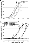
G50pKO virus reactivates from peritoneal exudate cells following intraperitoneal infection. Female C57BL/6 mice 6 to 8 weeks of age were infected with 1,000 PFU of G50pKO or G50pKO.MR virus by intraperitoneal injection. Splenocytes and PECs were harvested at day 18 following infection and assessed for establishment of and reactivation from latency by limiting dilution assays. (A) Splenocytes and PECs were plated in serial dilutions and subjected to nested PCR to detect G50. The percentage of genome-positive cells in each dilution was used to calculate the frequency of genome-positive cells as in reference . (B) Splenocytes and PECs were plated in serial dilutions onto MEF monolayers as in Fig. 9. The percentage of wells exhibiting cytopathic effect in each dilution was used to calculate the frequency of cells reactivating from latency. Mechanically disrupted cells were plated in parallel for each virus and cell type and verified the absence of preformed infectious virus (data not shown). Data are representative of at least two independent experiments with at least four mice per group, and error bars were generated using the standard errors of the means.

Similar articles

Cited by

References

    1. Allen, R. D., III, S. Dickerson, and S. H. Speck. 2006. Identification of spliced gammaherpesvirus 68 LANA and v-cyclin transcripts and analysis of their expression in vivo during latent infection. J. Virol. 802055-2062. - PMC - PubMed
    1. Ben-Sasson, S. A., and G. Klein. 1981. Activation of the Epstein-Barr virus genome by 5-aza-cytidine in latently infected human lymphoid lines. Int. J. Cancer 28131-135. - PubMed
    1. Bhende, P. M., W. T. Seaman, H. J. Delecluse, and S. C. Kenney. 2005. BZLF1 activation of the methylated form of the BRLF1 immediate-early promoter is regulated by BZLF1 residue 186. J. Virol. 797338-7348. - PMC - PubMed
    1. Bhende, P. M., W. T. Seaman, H. J. Delecluse, and S. C. Kenney. 2004. The EBV lytic switch protein, Z, preferentially binds to and activates the methylated viral genome. Nat. Genet. 361099-1104. - PubMed
    1. Blaskovic, D., M. Stancekova, J. Svobodova, and J. Mistrikova. 1980. Isolation of five strains of herpesviruses from two species of free living small rodents. Acta Virol. 24468. - PubMed

MeSH terms

LinkOut - more resources