. 2009 Feb;85(2):243-50.
doi: 10.1189/jlb.0608374. Epub 2008 Nov 6.

LPS activation is required for migratory activity and antigen presentation by tolerogenic dendritic cells

Affiliations

LPS activation is required for migratory activity and antigen presentation by tolerogenic dendritic cells

Amy E Anderson et al. J Leukoc Biol. 2009 Feb.

Abstract

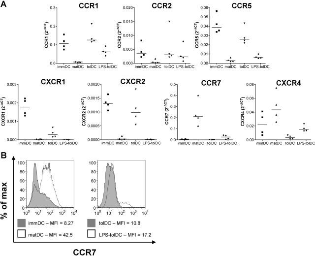
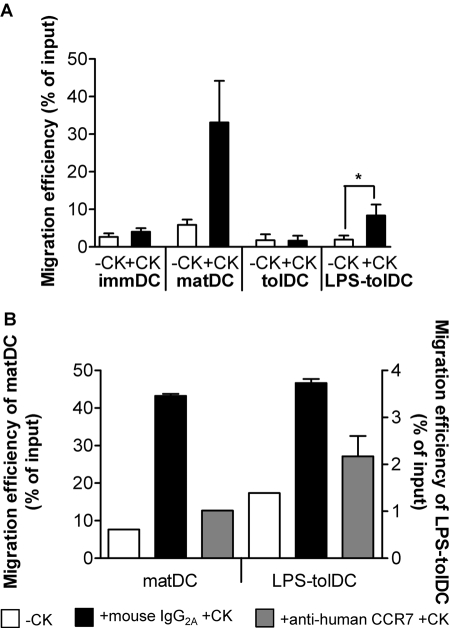
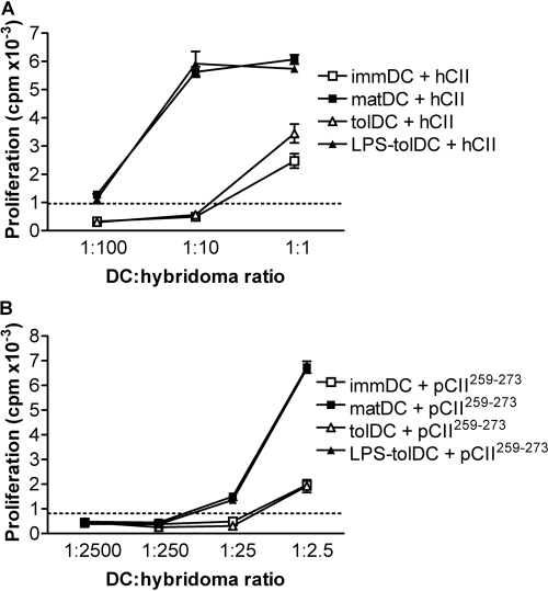
Autoimmune pathologies are caused by a breakdown in self-tolerance. Tolerogenic dendritic cells (tolDC) are a promising immunotherapeutic tool for restoring self-tolerance in an antigen-specific manner. Studies about tolDC have focused largely on generating stable maturation-resistant DC, but few have fully addressed questions about the antigen-presenting and migratory capacities of these cells, prerequisites for successful immunotherapy. Here, we investigated whether human tolDC, generated with dexamethasone and the active form of vitamin D3, maintained their tolerogenic function upon activation with LPS (LPS-tolDC), while acquiring the ability to present exogenous autoantigen and to migrate in response to the CCR7 ligand CCL19. LPS activation led to important changes in the tolDC phenotype and function. LPS-tolDC, but not tolDC, expressed the chemokine receptor CCR7 and migrated in response to CCL19. Furthermore, LPS-tolDC were superior to tolDC in their ability to present type II collagen, a candidate autoantigen in rheumatoid arthritis. tolDC and LPS-tolDC had low stimulatory capacity for allogeneic, naïve T cells and skewed T cell polarization toward an anti-inflammatory phenotype, although LPS-tolDC induced significantly higher levels of IL-10 production by T cells. Our finding that LPS activation is essential for inducing migratory and antigen-presenting activity in tolDC is important for optimizing their therapeutic potential.


Figures

Fig. 1.
Fig. 1.
LPS activation induces a semi-mature phenotype in tolDC with retention of an anti-inflammatory cytokine profile. (A) immDC, matDC, tolDC, and LPS-tolDC were stained with antibodies against cell-surface molecules, as indicated, and were assessed by flow cytometry. Debris and dead cells were excluded on the basis of forward-scatter and side-scatter, and viability was detected with Via-Probe. One representative experiment of 12 independent donors is shown. (B) DC populations were washed, cultured at 1.5 × 10⁵ DC/ml, and activated with CD40L-expressing cells (1.5×10⁵/ml) for 24 h. Cytokines in supernatants were quantified by sandwich ELISA. Error bars represent sem of triplicates. One representative experiment of five independent donors is shown. Detection level of IL-12p70 ELISA was 30 pg/ml.
Fig. 2.
Fig. 2.
tolDC and LPS-tolDC have differential expression of chemokine receptors. (A) Expression of chemokine receptor genes by DC was measured using a custom Micro Fluidic Card (Applied Biosystems). mRNA expression was normalized to that of human GAPDH by subtracting the CT value of the gene of interest from the CT value of the human GAPDH gene (ΔCT). Results are expressed as 2–ΔCT for four independent experiments. Horizontal lines represent median values. (B) DC were harvested 48 h after the addition of LPS and/or Dex and VitD3. DC populations were stained with anti-human CCR7 and were assessed by flow cytometry. Debris and dead cells were excluded on the basis of forward-scatter and side-scatter. The median fluorescent intensity (MFI) is shown. One representative experiment of three independent donors is shown.
Fig. 3.
Fig. 3.
LPS activation of tolDC induces migratory activity in response to the CCR7 ligand CCL19. DC populations were left untreated (A) or were treated with 5 μg/ml anti-human CCR7 antibody or mouse IgG2A isotype control for 30 min prior to assessment of migration. (B) Migration of DC was measured over a 2 h period in a transwell system. –CK, No chemokine; +CK, CCL19 (250 ng/ml) in the lower compartment. Migration efficiency was calculated as the percentage of the input DC that had migrated. Data are shown as the mean ± sem of five independent experiments (A) or are representative of three independent experiments; mean ± sem of duplicates are shown (B). * P < 0.05.
Fig. 4.
Fig. 4.
LPS activation of tolDC promotes antigen presentation. DC, generated from a HLA-DR1+ donor, were loaded with 20 μg/ml hCII (A) or 20 μg/ml nonglycosylated peptide pCII259–273 (B) on day 6, along with the addition of LPS and/or Dex and VitD3, or were left unloaded. After 24 h, DC were washed and cocultured with the T cell hybridoma HCII-9.1, which is specific for hCII259–273 (ratios are indicated in the figure). IL-2 production by the T cell hybridomas was measured by bioassay as the proliferative response of the IL-2-responsive cell line, CTLL-2. Proliferation was determined during the last 18 h of culture by incorporation of 3H-thymidine. Results are representative of two independent experiments. The dotted line shows the maximum background proliferation induced by unloaded DC. Error bars represent sem of triplicates.
Fig. 5.
Fig. 5.
tolDC and LPS-tolDC have low stimulatory capacity and similar immunomodulatory effects on allogeneic, naïve T cells. (A) matDC, tolDC, or LPS-tolDC (1×10⁴ cells/well) were cocultured with allogeneic, naïve T cells (1×10⁵ cells/well). Proliferation, measured by 3H-thymidine uptake, and IFN-γ production were assessed at day 6. Results are representative of eight independent experiments. Error bars represent sem of triplicates. (B–D) Allogeneic, naïve T cells were primed with DC (one DC:10 T cells) for 6 days and rested for 4 days with 0.1 ng/ml rIL-2. T cell lines primed by matDC (Tmat), tolDC (Ttol), and LPS-tolDC (TLPS-tol) were restimulated with matDC (B) or CD3/CD28 expander beads (C). Proliferation was assessed on day 3 by 3H-thymidine incorporation, and cytokine levels were measured by sandwich ELISA. Results of 10 (B) and 14 (C) independent experiments are shown. Horizontal lines represent mean values. (D) Primed T cells were restimulated with CD3/CD28 expander beads for 18 h, and IL-10 production was assessed using an IL-10 secretion assay. A CD3+CD4+ gate, identifying T cells, was used to exclude DC from subsequent analysis. The cytokine-positive gates shown were determined using unstimulated Tmat, Ttol, and TLPS-tol populations. Results are representative of two independent experiments. IL-10 APC, IL-10 allophycocyanin. (E) The mean production of IL-10 was divided by the mean production of IFN-γ (n=14; data from Fig. 4D), and the ratio was normalized to the IL-10:IFN-γ ratio of Tmat, which was set to 1. *, P < 0.05; **, P < 0.01.

References

    1. Lutz M B, Schuler G. Immature, semi-mature and fully mature dendritic cells: which signals induce tolerance or immunity? Trends Immunol. 2002;23:445–449. - PubMed
    1. Mahnke K, Schmitt E, Bonifaz L, Enk A H, Jonuleit H. Immature, but not inactive: the tolerogenic function of immature dendritic cells. Immunol Cell Biol. 2002;80:477–483. - PubMed
    1. Steinman R M, Hawiger D, Nussenzweig M C. Tolerogenic dendritic cells. Annu Rev Immunol. 2003;21:685–711. - PubMed
    1. Moser M. Dendritic cells in immunity and tolerance—do they display opposite functions? Immunity. 2003;19:5–8. - PubMed
    1. Gorczynski R M, Bransom J, Cattral M, Huang X, Lei J, Xiaorong L, Min W P, Wan Y, Gauldie J. Synergy in induction of increased renal allograft survival after portal vein infusion of dendritic cells transduced to express TGFβ and IL-10, along with administration of CHO cells expressing the regulatory molecule OX-2. Clin Immunol. 2000;95:182–189. - PubMed

Publication types

MeSH terms