. 2009 Jan;20(1):338-47.
doi: 10.1091/mbc.e08-04-0372. Epub 2008 Oct 29.

Multiple regulatory steps control mammalian nonmuscle myosin II assembly in live cells

Affiliations

Multiple regulatory steps control mammalian nonmuscle myosin II assembly in live cells

Mark T Breckenridge et al. Mol Biol Cell. 2009 Jan.

Abstract

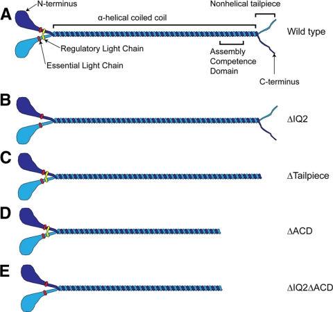
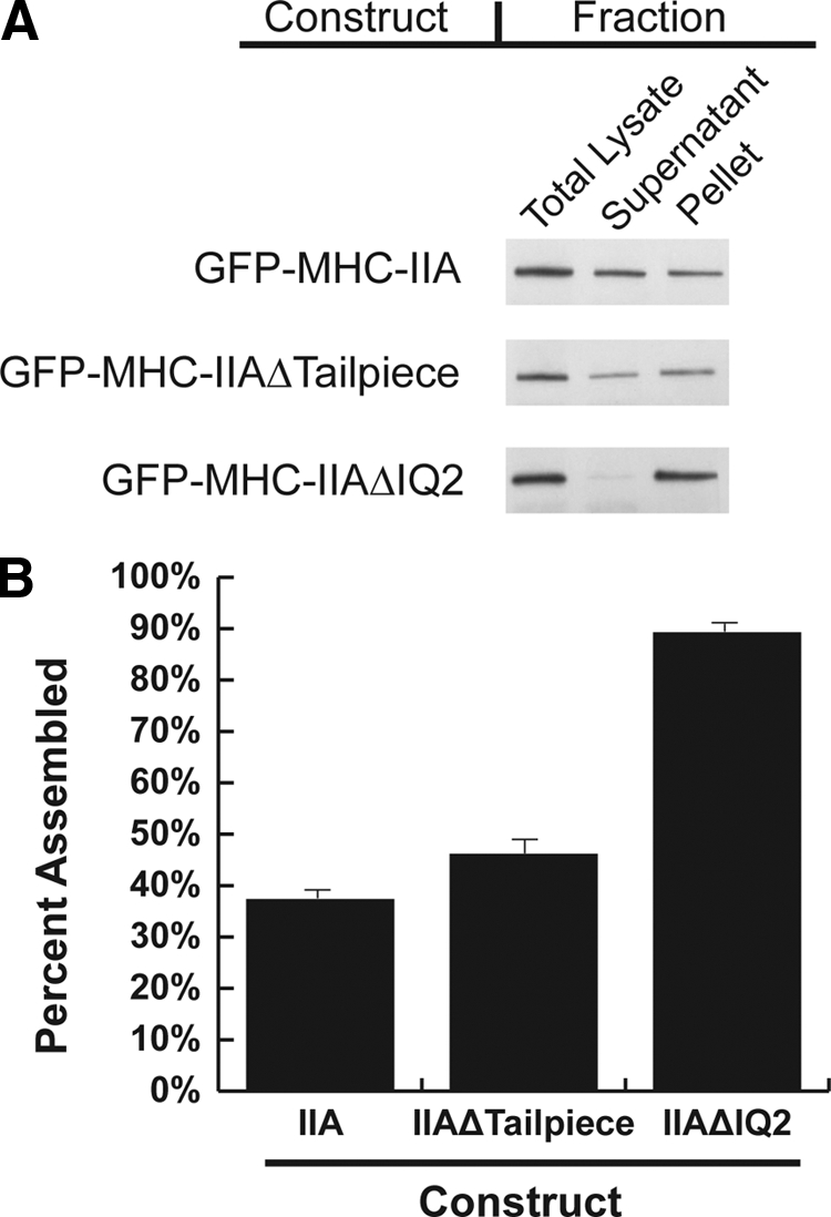
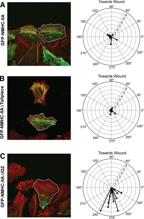
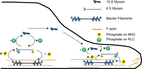
To better understand the mechanism controlling nonmuscle myosin II (NM-II) assembly in mammalian cells, mutant NM-IIA constructs were created to allow tests in live cells of two widely studied models for filament assembly control. A GFP-NM-IIA construct lacking the RLC binding domain (DeltaIQ2) destabilizes the 10S sequestered monomer state and results in a severe defect in recycling monomers during spreading, and from the posterior to the leading edge during polarized migration. A GFP-NM-IIA construct lacking the nonhelical tailpiece (Deltatailpiece) is competent for leading edge assembly, but overassembles, suggesting defects in disassembly from lamellae subsequent to initial recruitment. The Deltatailpiece phenotype was recapitulated by a GFP-NM-IIA construct carrying a mutation in a mapped tailpiece phosphorylation site (S1943A), validating the importance of the tailpiece and tailpiece phosphorylation in normal lamellar myosin II assembly control. These results demonstrate that both the 6S/10S conformational change and the tailpiece contribute to the localization and assembly of myosin II in mammalian cells. This work furthermore offers cellular insights that help explain platelet and leukocyte defects associated with R1933-stop alleles of patients afflicted with human MYH9-related disorder.


Figures

Figure 1.
Figure 1.
GFP fusion constructs used to assess myosin IIA assembly models. (A) Wild-type NM-II. (B) The NM-II with the IQ2 region deleted, preventing RLC binding. (C) The NM-II with the nonhelical tailpiece deleted. (D) The NM-II with the assembly competence domain (ACD) deleted. The ACD is required for assembly of the NM-II monomer into bipolar filaments. (E) The NM-II with the ACD deleted and the IQ2 region deleted.
Figure 2.
Figure 2.
GFP-NM-IIAΔIQ2ΔACD displays reduced diffusional rates in live cells consistent with destabilization of the 10S folded state. (A) Western blots of immunoprecipitations depicting RLC association with each NM-II. (B) Images depict representative cells transfected with indicated assembly incompetent myosin construct. The live cell diffusional characteristics of each myosin was then measured via FRAP analysis. Plots show average recovery for all experiments (n = 90) plotted in solid black with ±SD shown in dashed gray. Deleting the RLC-binding site increased the half-time of recovery 1.23-fold compared with GFP-NM-IIAΔACD. GFP-NM-IIAΔACD had a half-time of recovery of 0.96 ± 0.45 s. GFP-NM-IIAΔIQ2ΔACD had a significantly slower half-time of recovery of 1.18 ± 0.53 s (p = 0.003). Average half times ± SD. p values were determined by two-tailed t test. Scale bars, 10 μm.
Figure 3.
Figure 3.
In high-speed cleared cell lysates, GFP-NM-IIAΔIQ2 displayed dramatically greater assembly than did GFP-NM-IIA at 140 mM salt. Essentially all of the GFP-NM-IIA is soluble at 140 mM and at 400 mM salt. However, at 140 mM salt a substantial portion of the GFP-NM-IIAΔIQ2 is assembled and pellets. However, this effect was eliminated at 400 mM salt. This result strongly suggests that the ΔIQ2 mutation causes the myosin to remain filamentous by destabilizing the 10S folded species. Western blot was probed with a GFP antibody.
Figure 4.
Figure 4.
The NM-IIAΔIQ2 and NM-IIAΔtailpiece recombinant proteins can assemble into stress fibers in undisturbed cultured cells. Cells were transiently transfected with GFP-NMHC-IIA constructs and stained with Alexa Fluor 568 phalloidin (actin) and DAPI (DNA stain). Scale bars, 10 μm.
Figure 5.
Figure 5.
Both NM-IIAΔIQ2 and NM-IIAΔtailpiece display over-assembly in a cytoskeletal ghost assay. (A) Western blots of Triton-insoluble fractions of cells transfected with the indicated construct probed with an anti-GFP antibody. Equivalent fractions were loaded based on cell concentration. (B) Densitometry was performed on blots from seven to eight separate experiments. Error bars, SEs; n = 7 or 8.
Figure 6.
Figure 6.
Altered lamellar localization and assembly of mutant GFP-NM-IIA constructs. Actively spreading cells transfected with the indicated construct were fixed and stained for actin. The pixel intensity values for all equidistant pixels from the cell boundary were averaged for both the myosin image and actin image. Average pixel intensity was then plotted as a function of distance from the cell edge. Distances were normalized to the maximum distance from the cell edge. Therefore, 0% on the x-axis is the cell edge, and 100% is the maximum distance from the cell edge. Images and plots from a typical cell are shown for each construct. For pooled data, see Table 1. Scale bars, 10 μm. Relative to the wild-type GFP-NM-IIA construct (A), the Δtailpiece construct (B), and S1943A construct (D), are recruited to the spreading lamella and display marginal over-assembly relative to the cell center (Table 1). In contrast, GFP-NM-IIAΔIQ2 (C) displayed severe over-assembly in the cell center, and was excluded from the spreading margin.
Figure 7.
Figure 7.
NM-IIAΔIQ2 displays a severe defect in recycling to the leading edge of polarized migrating cells. Cells transfected with the indicated myosin construct (green) were fixed during polarized migration and stained for actin (red). Individual cells from images of the wound margin were outlined by hand and their centroid of GFP intensity relative to the center of cell geometry was determined. The left panels show a typical field of cells with a traced cell (outlined in white) used for analysis. Scale bars, 10 μm. The right panels show compass plots with vectors calculated from all experiments. On the compass plots, 90° is toward the wound, the radial axis is percent of cell perimeter and zero is the center of cell geometry. (A) GFP-NM-IIA exhibits a close localization to the center of cell shape, with a mild forward bias (0.85 ± 0.15% at 27°, n = 11). (B) GFP-NM-IIAΔtailpiece exhibited a mild, but consistent bias behind the geometrical centroid (0.51 ± 0.11% at 318° for IIAΔtailpiece, n = 12), suggesting a possible assembly/disassembly control defect. (C) GFP-NM-IIAΔIQ2 exhibited a pronounced and consistent rearward shift (3.41 ± 0.53% at 284°, n = 11).
Figure 8.
Figure 8.
FRAP dynamics at the lamellar margin of spreading cells reveals assembly defects for both NM-IIAΔIQ2 and NM-IIAΔtailpiece. FRAP was performed on GFP-myosin assembled at the lamella of actively spreading cells transfected with the indicated construct. Images depict examples of a typical cell transfected with the indicated construct with its associated recovery curve. Scale bars, 10 μm. The Δtailpiece had a similar recovery to wild type, whereas the ΔIQ2 actually had faster halftime (Table 2). Both Δtailpiece and ΔIQ2 had larger immobile fractions compared with wild type, suggesting that they both are biased toward an assembled state.
Figure 9.
Figure 9.
Model for in vivo control of myosin IIA assembly in mammalian nonmuscle cells. In this model, sequestration in the 10S form plays important roles in maintaining the normal equilibrium between assembly and disassembled states in live cells. Destabilization of the 10S species by the ΔIQ mutant dramatically shifts the in vivo equilibrium of monomer versus assembled pools toward the assembled state. This in turn severely compromises recycling to leading edge protrusions, which in wild type NM-II would be dependent on RLC dephosphorylation. On the basis of our data and other recent studies (see Discussion), we propose that regulation via the tailpiece specifically plays a role in driving the rapid local removal of myosin IIA subunits from assembled filaments, back to the linear, monomeric, 6S state. We suggest that the fundamental role of the tailpiece in this process is that it serves as a target for phosphorylation at S1943.

References

    1. Adelstein R. S., Pato M. D., Conti M. A. The role of phosphorylation in regulating contractile proteins. Adv. Cyclic. Nucleotide Res. 1981;14:361–373. - PubMed
    1. Bao J., Jana S. S., Adelstein R. S. Vertebrate nonmuscle myosin II isoforms rescue small interfering RNA-induced defects in COS-7 cell cytokinesis. J. Biol. Chem. 2005;280:19594–19599. - PubMed
    1. Bray D., White J. G. Cortical flow in animal cells. Science. 1988;239:883–888. - PubMed
    1. Burgess S. A., Yu S., Walker M. L., Hawkins R. J., Chalovich J. M., Knight P. J. Structures of smooth muscle myosin and heavy meromyosin in the folded, shutdown state. J. Mol. Biol. 2007;372:1165–1178. - PubMed
    1. Conti M. A., Adelstein R. S. Nonmuscle myosin II moves in new directions. J. Cell Sci. 2008;121:11–18. - PubMed

Publication types