Comparative Study
. 2008 Oct 29;28(44):11263-8.
doi: 10.1523/JNEUROSCI.2308-08.2008.

Up-regulation of P2X4 receptors in spinal microglia after peripheral nerve injury mediates BDNF release and neuropathic pain

Affiliations
Comparative Study

Up-regulation of P2X4 receptors in spinal microglia after peripheral nerve injury mediates BDNF release and neuropathic pain

Lauriane Ulmann et al. J Neurosci. .

Abstract

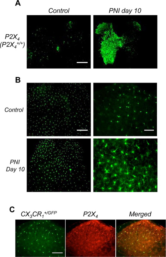
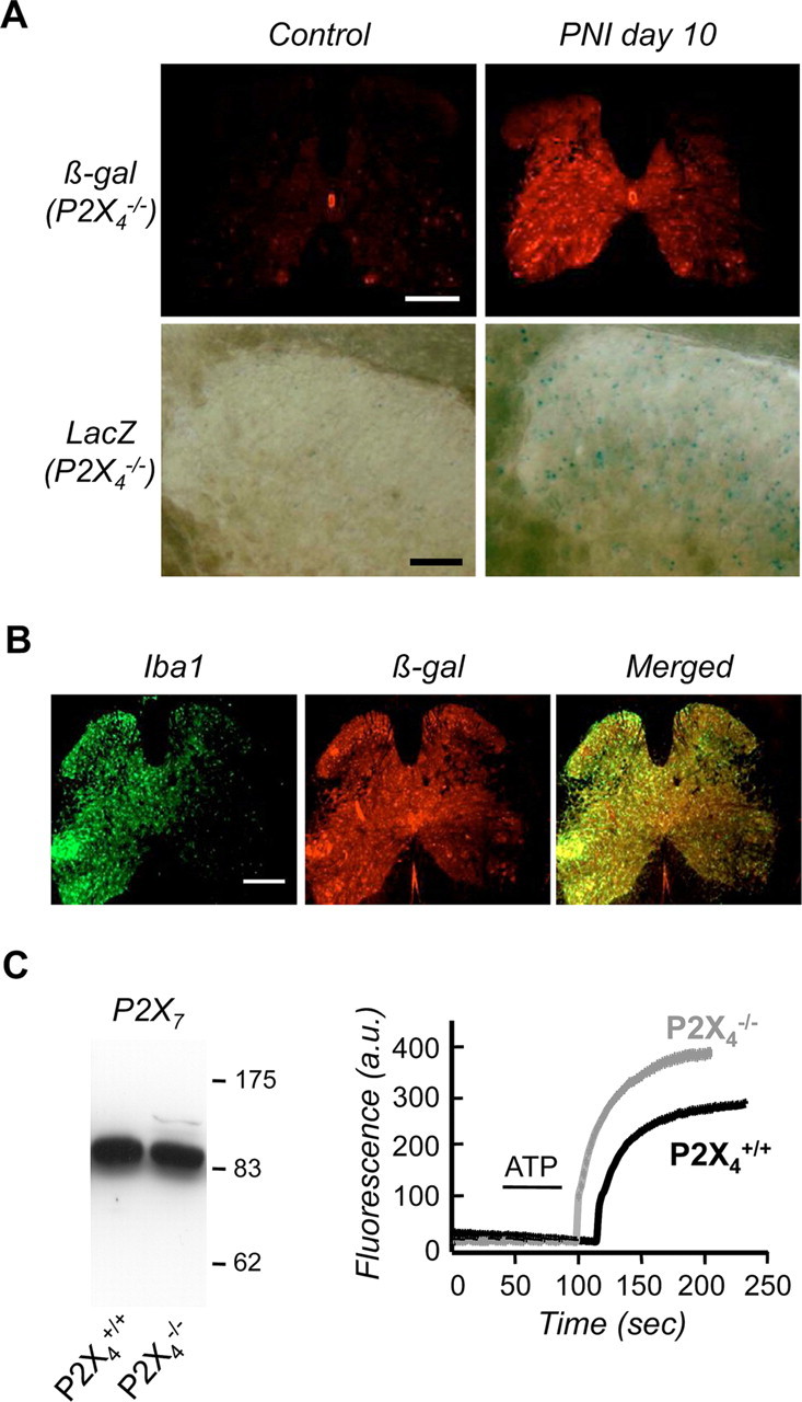
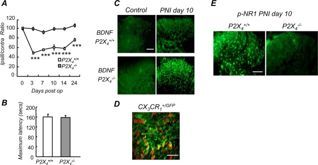
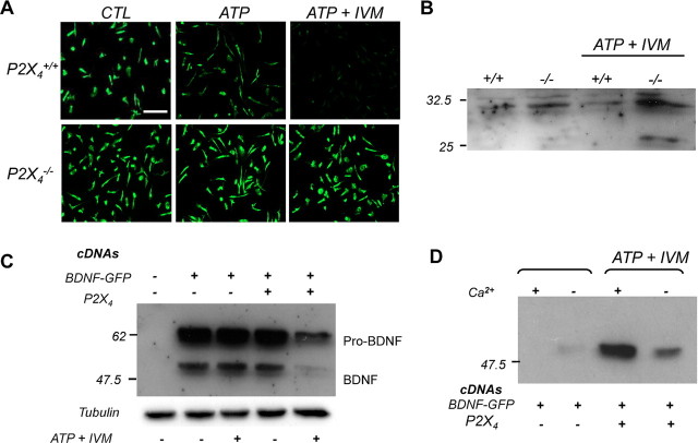
ATP is a known mediator of inflammatory and neuropathic pain. However, the mechanisms by which specific purinergic receptors contribute to chronic pain states are still poorly characterized. Here, we demonstrate that in response to peripheral nerve injury, P2X(4) receptors (P2X(4)R) are expressed de novo by activated microglia in the spinal cord. Using in vitro and in vivo models, we provide direct evidence that P2X(4)R stimulation leads to the release of BDNF from activated microglia and, most likely phosphorylation of the NR1 subunit of NMDA receptors in dorsal horn neurons of the spinal cord. Consistent with these findings, P2X4-deficient mice lack mechanical hyperalgesia induced by peripheral nerve injury and display impaired BDNF signaling in the spinal cord. Furthermore, ATP stimulation is unable to stimulate BDNF release from P2X(4)-deficient mice microglia in primary cultures. These results indicate that P2X(4)R contribute to chronic pain through a central inflammatory pathway. P2X(4)R might thus represent a potential therapeutic target to limit microglia-mediated inflammatory responses associated with brain injury and neurodegenerative disorders.


Figures

Figure 1.
Figure 1.
Peripheral nerve injury induces upregulation of P2X4R in activated spinal microglia. A, Comparison of P2X4 expression in the spinal cord in sham-operated and injured mice. P2X4 immunoreactivity was barely detectable in sham animals whereas 10 d PNI, it was clearly induced ipsilateral to the lesion. Note that P2X4 expression spread to the whole side of the spinal cord because of sensory and motor fiber lesions. Scale bar, 500 μm. B, Morphology of spinal microglia in CX3CR1+/GFP mice post-PNI. Compared with sham animals, 10 d post-PNI microglia displayed higher fluorescence intensities, whereas the number of GFP cells was not significantly different. Increase of fluorescence intensity was restricted to the ipsilateral side of the lesion. Scale bar, 500 μm. At higher magnification in the dorsal horn region of the spinal cord (right), post-PNI microglia presented the typical morphological characteristics of the activated state with larger cell bodies and compacted processes compared with microglia from sham animals. Scale bar, 50 μm. C, PNI induces P2X4 expression in activated microglia. In the dorsal horn, P2X4 immunoreactivity colocalized exclusively with microglia-specific eGFP fluorescence in CX3CR1+/GFP mice 10 d post-PNI. Scale bar, 100 μm.
Figure 2.
Figure 2.
Microglial upregulation of P2X4 gene expression induced by PNI is still present in P2X4-deficient mice. A, In P2X4−/− mice, PNI induced β-galactosidase expression in the spinal cord ipsilateral to the lesion whereas no expression was detected in sham animals. Top row, β-Galactosidase immunoreactivity in spinal cord; bottom row: LacZ staining in the dorsal horn (scale bar, 500 μm). B, Post-PNI, ß-galactosidase immunoreactivity (red) colocalized with the microglial marker Iba1 (green). Scale bar, 500 μm. C, P2X4-gene deletion did not affect P2X7 expression and function in microglia. Western blot of P2X7 expression (left) and ATP (1 mm) induced YOPRO uptake in primary microglial culture from wild-type and P2X4−/− mice (right). Representative experiment of 3 showing mean fluorescence intensity of all recorded cells (>50) after background substraction.
Figure 3.
Figure 3.
Lack of mechanical hypersensitivity and altered BDNF signaling in P2X4-deficient mice after peripheral nerve injury. A, Time course of mechanical hypersensitivity induced by sciatic nerve ligature in wild-type and P2X4−/− mice expressed as ipsilateral/contralateral ratios. No hypersensitivity was observed in the P2X4−/− mice, whereas wild-type mice develop a strong hypersensitivity lasting >23 d, (n = 15 mice, two-way ANOVA, ***p < 0.005). B, Sensorimotor coordination was measured using the rotarod test. No difference between wild-type and P2X4−/− mice was observed, ruling out a potential motor deficit in the P2X4−/− mice. C, BDNF immunoreactivity in the dorsal horn of the spinal cord was analyzed by immunohistochemistry in wild-type and P2X4−/− mice. In sham-operated mice, BDNF immunoreactivity was not detected in either wild-type or P2X4−/− animals (left). Ten days post-PNI (PNI day 10), BDNF immunoreactivity was slightly increased in wild-type mice, whereas a strong immunoreactivity was observed in the P2X4−/− mice. Scale bar, 100 μm. D, In the dorsal horn of CX3CR1+/GFP mice 10 d post-PNI, BDNF immunoreactivity (red) colocalized with microglial eGFP fluorescence (green). Scale bar, 50 μm. E, PNI-induced phosphorylation of the NR1 subunit in dorsal horn neurons was impaired in P2X4−/− mice. Levels of phosphorylation of NR1 were analyzed by immunohistochemistry. Scale bar, 100 μm.
Figure 4.
Figure 4.
P2X4 stimulation triggers BDNF release from activated microglia. A, ATP-evoked release of BDNF from wild-type and P2X4−/− microglia primary cultures. Intracellular BDNF content of microglia was analyzed by immunostaining. Stimulation of a culture with ATP (100 μm) alone or coapplied with ivermectin (IVM, 3 μm) induced a strong decrease of BDNF immunoreactivity in a primary microglial culture from wild-type mice. In a culture from P2X4−/− mice, ATP or ATP+IVM stimulation did not induce any detectable changes in BDNF immunoreactivity. The data shown are representative of six experiments. Scale bar, 100 μm. B, Western blot of intracellular BDNF content from primary cultures of wild-type and P2X4−/− microglia. Cells were stimulated as above. The top band corresponds to pro-BDNF; the mature form (i.e., the 14 kDa form) could not be resolved in these experiments. ATP+IVM stimulation induced a reduction of intracellular pro-BDNF in wild-type but not in P2X4−/− cultures. The intermediate form seen in stimulated P2X4−/− cultures is of unknown origin. C, D, P2X4 activation triggered BDNF release in a recombinant expression system. P2X4 and BDNF-GFP cDNAs were transiently transfected in COS-7 cells either alone or in combination. Forty-eight hours later, cells were stimulated or not with ATP+IVM and intracellular (C) or secreted (D) BDNF-GFP was analyzed by Western blotting. Note that in the absence of extracellular Ca2+, ATP-induced secretion of BDNF was strongly reduced.

References

    1. Biber K, Neumann H, Inoue K, Boddeke HW. Neuronal ‘On’ and ‘Off’ signals control microglia. Trends Neurosci. 2007;30:596–602. - PubMed
    1. Buell G, Michel AD, Lewis C, Collo G, Humphrey PP, Surprenant A. P2X1 receptor activation in HL60 cells. Blood. 1996;87:2659–2664. - PubMed
    1. Burnstock G. Physiology and pathophysiology of purinergic neurotransmission. Physiol Rev. 2007;87:659–797. - PubMed
    1. Chessell IP, Hatcher JP, Bountra C, Michel AD, Hughes JP, Green P, Egerton J, Murfin M, Richardson J, Peck WL, Grahames CB, Casula MA, Yiangou Y, Birch R, Anand P, Buell GN. Disruption of the P2X7 purinoceptor gene abolishes chronic inflammatory and neuropathic pain. Pain. 2005;114:386–396. - PubMed
    1. Coull JA, Boudreau D, Bachand K, Prescott SA, Nault F, Sík A, De Koninck P, De Koninck Y. Trans-synaptic shift in anion gradient in spinal lamina I neurons as a mechanism of neuropathic pain. Nature. 2003;424:938–942. - PubMed

Publication types

MeSH terms

Substances