Comparative Study
. 2008 Oct 29;28(44):11409-20.
doi: 10.1523/JNEUROSCI.2135-08.2008.

Requirement of 3-phosphoinositide-dependent protein kinase-1 for BDNF-mediated neuronal survival

Affiliations
Comparative Study

Requirement of 3-phosphoinositide-dependent protein kinase-1 for BDNF-mediated neuronal survival

Giorgi Kharebava et al. J Neurosci. .

Abstract

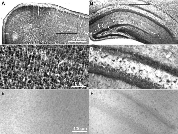
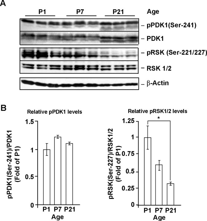
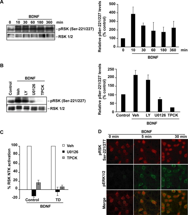
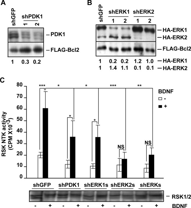
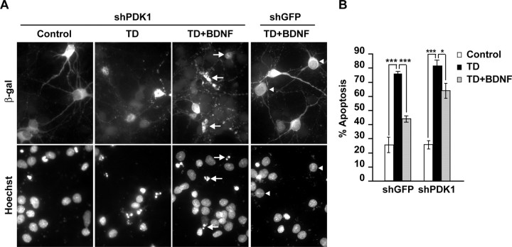
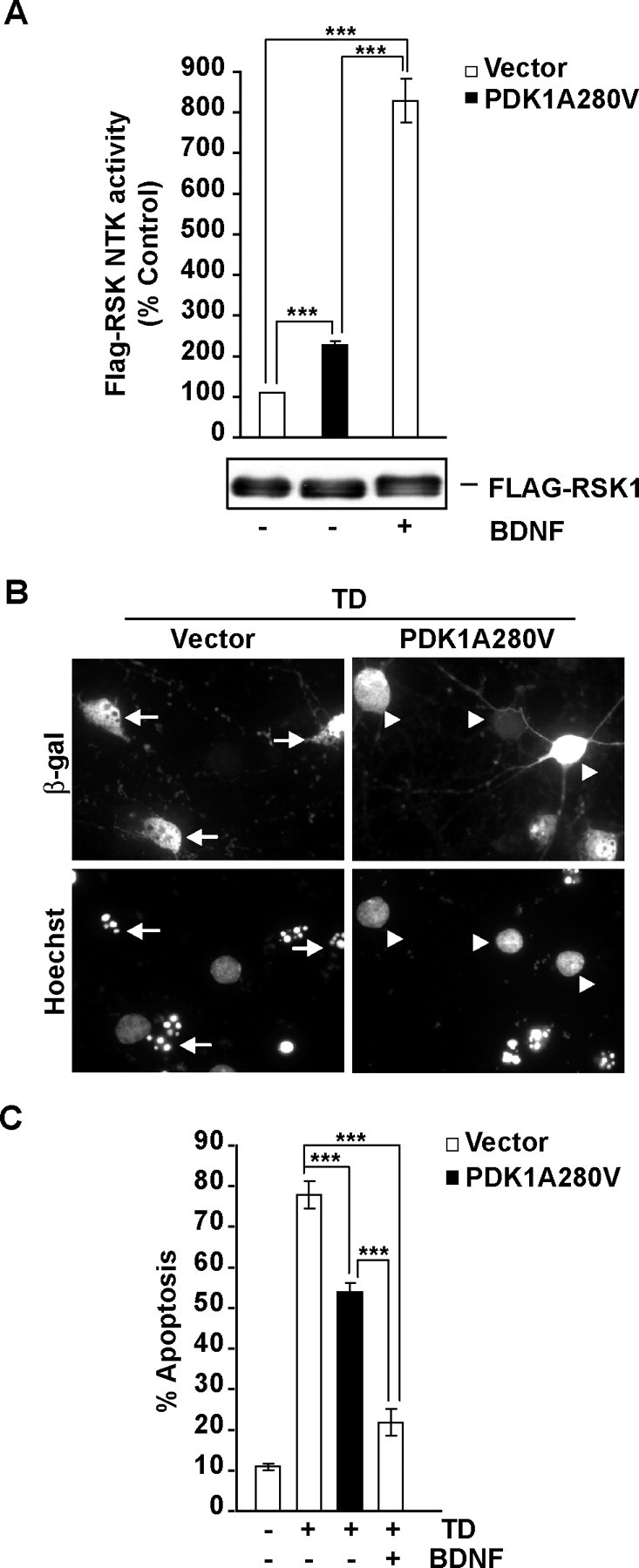
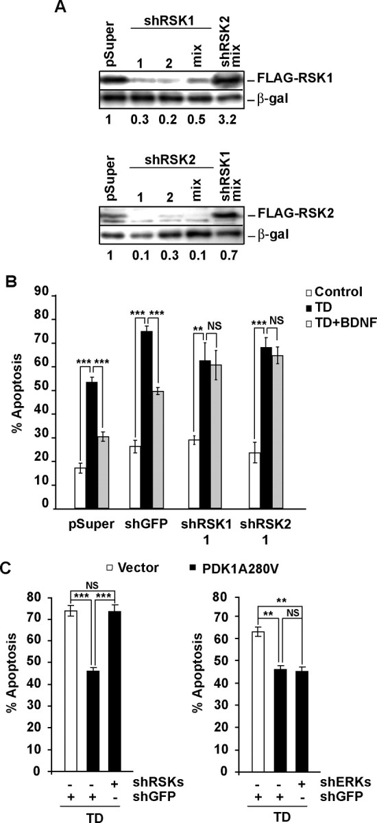
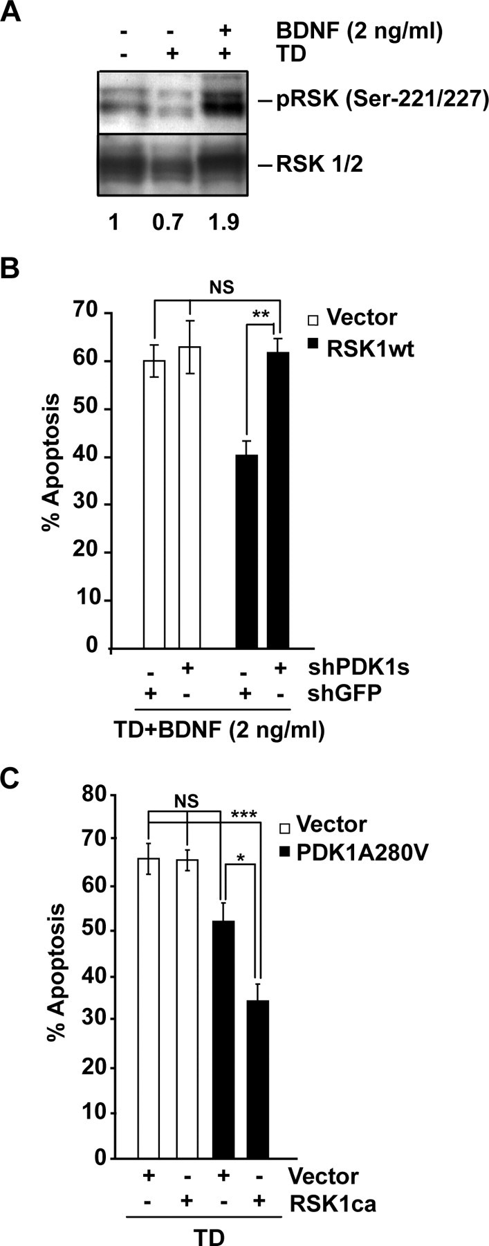
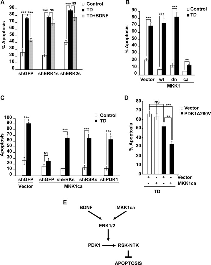
Although PDK1 regulates several signaling pathways that respond to neurotrophins, direct evidence for its involvement in neurotrophin-mediated survival has not yet been reported. Here we show high neuronal expression of active PDK1 in the rat cortex and hippocampus at the developmental stages with pronounced dependence on extracellular survival signals. Also, in cultured cortical neurons from newborn rats, BDNF resulted in PDK1- and extracellular signal-regulated kinase-1/2 (ERK1/2)-mediated activation of their direct target, the p90 ribosomal S6 kinase 1/2 (RSK1/2). In trophic-deprived cortical neurons, knockdown of endogenous PDK1 attenuated the antiapoptotic survival response to 10 ng/ml BDNF, whereas an overexpressed active mutant form of PDK1 reduced apoptosis. The neuroprotection by BDNF or active PDK1 required RSK1/2. Conversely, PDK1 knockdown reversed the survival effects of combining the overexpressed RSK1 with a low, subprotective BDNF concentration of 2 ng/ml. Likewise, the protection by the overexpressed, active PDK1 was enhanced by coexpression of an active RSK1 mutant. Consistent with the observations that in BDNF-stimulated neurons RSK1/2 activation required both PDK1 and ERK1/2, ERK1/2 knockdown removed BDNF-mediated survival. Selective activation of ERK1/2 with an overexpressed active mutant form of MKK1 resulted in RSK1/2- and PDK1-dependent neuroprotection. Finally, at subprotective plasmid DNA dosage, overexpression of the active MKK1 and PDK1 mutants produced synergistic effect on survival. Our findings indicate a critical role for PDK1-RSK1/2 signaling in BDNF-mediated neuronal survival. Thus, the PDK1 is indispensable for the antiapoptotic effects of the ERK1/2 pathway offering previously unrecognized layer of survival signal processing and integration.


Figures

Figure 1.
Figure 1.
PDK1 expression in the rat brain. A–D, PDK1 immunoreactivity in coronal sections through forebrains of rat pups at P7. PDK1 was visualized using ABC/DAB immunohistochemistry. E, F, The specificity of PDK1 labeling was confirmed in control experiments, in which the primary antibody was replaced by the nonimmune rabbit IgG. Intense PDK1 staining was found in neuron-like cells in the neocortical layers II–VI (A, C) as well as the pyramidal cell layer of the Ammon's horn and granule cell layer of the dentate gyrus (B, D). Similar pattern of PDK1 immunoreactivity was observed at P21 (data not shown). CA, Cornu amonis; DG, dentate gyrus.
Figure 2.
Figure 2.
Regulation of PDK1 during rat cortical development. A, B, At P1, P7, and P21, both total PDK1 and phospho-Ser-241 PDK1 were detected by Western blotting of lysates from rat cortex. Note that the pSer241-PDK1/total PDK1 ratio remained similar at all developmental stages examined. In contrast, the PDK1-mediated phosphorylation of RSK1/2 Ser221/227 decreased over the studied ages. Thus, PDK1-RSK1/2 signaling is active in developing forebrain during synaptogenesis when neurons are highly dependent on extracellular survival signals. At P1 and P7, electromobility of total or pSer241 PDK1 was lower than at P21, indicating possible hyperphosphorylation at non-Ser241 sites. In A, same amounts of protein lysates were analyzed in each lane. In B, data represent averages of three animals at each developmental stage ±SEM; the pSer241 PDK1 or pSer221/227 RSK1/2 levels were normalized against total PDK1 or RSK1/2, respectively. *p < 0.05.
Figure 3.
Figure 3.
PDK1-mediated activation of RSK1/2 in BDNF-stimulated neurons. Neurons were stimulated with 10 ng/ml BDNF as indicated. In A, B, Western blot analysis of RSK1/2 phosphorylation at Ser221/227 is presented. Blots were reprobed to detect total RSK1/2. For quantifications, the ratios of pSer221/227 to total RSK1/2 were compared with unstimulated controls; averages of two independent experiments ±SD are presented. A, BDNF increased PDK1-mediated phosphorylation of RSK1/2. B, The BDNF-mediated increase of pSer221/227 required activities of the ERK1/2 pathway and PDK1. Although in neurons treated with either the vehicle (Veh; 0.2% DMSO) or the 30 μm PI3K inhibitor LY294002 (LY), a 1 h BDNF treatment elevated pSer221/227, the 50 μm MKK1/2 inhibitor U0126 or the 50 μm PDK1 pathway inhibitor TPCK blocked that effect. Experiments providing additional validation of the inhibitors are presented in supplemental Figure S1B, available at www.jneurosci.org as supplemental material. C, Without drug inhibitors (Veh), cortical neurons that were sham treated (control) or trophic deprived for 3 h and then stimulated with BDNF for 1 h showed increased RSK1/2 NTK activity as determined by immunocomplex kinase assay. The ERK1/2 or PDK1 pathway inhibition abolished RSK1/2 NTK activation by BDNF. Data represent two independent experiments ±SD. D, The coincidence of the MKK1/2-ERK1/2 and PDK1-RSK1/2 signaling in BDNF stimulated neurons. Representative confocal images are of neurons that were coimmunostained for pERK1/2 (pThr183-pTyr185, green) and pSer221/227 (red). BDNF stimulations were performed 1 h after placing neurons in serum-free media supplemented with MK801 to reduce the basal ERK1/2 signaling. Note nuclear localization of PDK1-phosphorylated RSK1/2 in BDNF-treated neurons. Most pERK appeared cytosolic. Similar patterns were observed in two independent experiments. Additional data illustrating the coincidence of the BDNF-mediated RSK1/2 activations at the NTK and CTK are presented in supplemental Figure S2, available at www.jneurosci.org as supplemental material.
Figure 4.
Figure 4.
Knockdown of PDK1 reduces ERK-dependent activation of RSK1/2. A, Freshly isolated cortical neurons were coelectroporated with two different shRNA constructs targeting PDK1 (shPDK1-1 or -2) together with an expression plasmid for FLAG-Bcl2 that was added to reduce electroporation toxicity (4 + 0.2 μg of each plasmid DNA/5 × 106 cells, respectively). The shRNA against GFP was used as a control (shGFP). After 72 h, Western blot analysis revealed that shPDK1-1 and shPDK1-2 reduced the levels of endogenous PDK1 by 70 or 80% of control, respectively. Numbers under the blot represent the ratios of endogenous PDK1 to Flag-Bcl2 levels. B, Freshly isolated cortical neurons were coelectroporated with pcDNA3-HA-ERK1, pcDNA3-HA-ERK2, and pcDNA3-FLAG-Bcl2 together with one of the following: shGFP, shERK1-1, shERK1-2, shERK2-1, and shERK2-2, as indicated (0.8 + 0.8 + 0.2 + 2.5 μg of each plasmid DNA/5 × 106 neurons, respectively). After 72 h, the levels of HA-ERK1/2 were analyzed by Western blotting with an anti-HA antibody. Numbers under the blot represent the ratios of the indicated HA-ERKs to Flag-Bcl2 levels. Reduced levels of HA-ERK1 or HA-ERK2 compared with Flag-Bcl2 indicate efficient knockdowns with shERK1 or shERK2, respectively. C, Cortical neurons were electroporated with the indicated shRNAs (3 μg plasmid DNA/10 × 106 cells). To reduce electroporation toxicity, the dominant negative p53 expression plasmid (p53-DD) was included in all samples (1 μg plasmid DNA/10 × 106 cells). After 72 h, neurons were stimulated with BDNF for 30 min and the NTK activity of the endogenous RSK1/2 was analyzed by immunocomplex kinase assay. Immunoprecipitates contained similar levels of RSK1/2 as determined by Western blotting (shown at the bottom of the graph). Depletion of PDK1 or ERK1 reduced the BDNF activation of RSK NTK. Depletion of ERK2 abolished that activation. Therefore, ERK1, ERK2, and PDK1 are required for full activation of neuronal RSK1/2 by BDNF. In A, B, similar results were obtained in two independent experiments. In C, averages of three independent experiments ±SEM are presented. *p < 0.05; **p < 0.01; ***p < 0.001; nonsignificant (NS), p > 0.05.
Figure 5.
Figure 5.
Requirement of PDK1 for antiapoptotic activity of BDNF in trophic-deprived neurons. Neurons were cotransfected with shGFP or an equimolar mix of shPDK1-1 and shPDK1-2 (shPDK1) together with the β-galactosidase expression plasmid, pON260 (1 + 0.2 μg plasmid DNA/5 × 105 neurons, respectively). After 24 h, neurons were sham treated or trophic deprived in the presence of 0 or 10 ng/ml BDNF for the next 24 h. A, Representative photomicrographs illustrating morphology of the transfected neurons, which were identified by β-galactosidase immunostaining. Counterstaining with Hoechst 33258 revealed apoptotic alterations including condensation and fragmentation of the chromatin (arrows). Arrowheads point β-galactosidase-positive cells without signs of apoptosis. B, The knockdown of PDK1 reduced BDNF protection against TD-induced apoptosis. The individual shPDK1 plasmids affected BDNF survival similarly to their mix (data not shown). Averages of triplicate determinations from three independent experiments ±SEM are presented. *p < 0.05; ***p < 0.001.
Figure 6.
Figure 6.
Neuroprotection against the TD-induced apoptosis by the activated mutant form of PDK1, PDK1A280V. A, Cortical neurons were coelectroporated with Flag-RSK1 and either pcDNA3 (vector) or the activated PDK1 mutant PDK1A280V as indicated (3 + 3 μg/10 × 106 cells). At 17 h after electroporation, cells were stimulated with 0 (−) or 10 (+) ng/ml BDNF for 3 h. At 20 h after electroporation, the effects of PDK1A280V or BDNF were analyzed by immunocomplex kinase assay with the immunoprecipitated Flag-RSK1. Although similar levels of Flag-RSK1 were detected in all immunoprecipitates (as indicated by the anti-flag Western blot shown under the graph), BDNF treatment or PDK1A280V increased Flag-RSK1 NTK activity. B, C, Neurons were cotransfected with expression plasmids for β-galactosidase (EF1αLacZ) or the activated PDK1A280V mutant (0.2 + 1 μg of plasmid DNA/5 × 105 neurons, respectively). The empty expression vector pcDNA3 was used as a control (Vector). After 24 h, neurons were sham treated (control) or trophic deprived with or without 10 ng/ml BDNF, as indicated. Apoptosis was evaluated after the next 24 h. B, Representative photomicrographs of the transfected neurons, which were detected by β-galactosidase immunoreactivity. Transfected neurons with normal chromatin morphology or its apoptotic rearrangements are pointed by arrowheads or arrows, respectively. C, Compared with BDNF, the overexpressed PDK1A280V offered partial protection against TD-induced apoptosis. In A and C, averages of three independent experiments ±SEM are presented. ***p < 0.001.
Figure 7.
Figure 7.
RSK1/2 is required for PDK1-mediated neuroprotection. A, Freshly isolated cortical neurons were coelectroporated with indicated shRNA constructs, together with expression plasmids for β-galactosidase (EF1αLacZ) and either FLAG-RSK1 or FLAG-RSK2 as indicated (2.5 + 0.2 + 0.3 μg of each plasmid DNA/5 × 106 neurons, respectively). If mixed, shRNA plasmids were combined eqimolarly and applied at 2.5 μg of total plasmid DNA/5 × 106 neurons. The controls included the shRNA vector pSuper and the shRSK2 or shRSK1 mixes for shRSK1 or shRSK2, respectively. After 72 h, the levels of FLAG-RSK1 or FLAG-RSK2 but not β-galactosidase decreased in response to isoform-specific shRNAs indicating efficient and selective knockdown. B, Neurons were cotransfected with the β-galactosidase plasmid (pON260) and the indicated shRNAs (0.2 + 0.5 μg of plasmid DNA/0.5 × 106 neurons, respectively). After 48 h, neurons were sham- or TD-treated with or without 10 ng/ml BDNF for additional 24 h. In neurons receiving control plasmids (pSuper or shGFP), BDNF reduced the apoptotic responses to TD. The protection against TD was removed by knocking down either RSK1 or RSK2 indicating that each of these RSK isoforms was required for BDNF to suppress apoptosis. C, Neurons were cotransfected with pON260, the indicated shRNAs, and the activated PDK1A280V mutant (0.2 + 0.2 + 0.8 μg of plasmid DNA/0.5 × 106 neurons, respectively). After 24 h, neurons were trophic deprived for the next 24 h. PDK1A280V reduced TD-induced apoptosis in neurons receiving shGFP, whereas it failed to protect the recipients of the equimolar mix of shRSK1-1, shRSK1-2, shRSK2-1, and shRSK2-2 (shRSKs). In contrast, the equimolar mix of shERK1-1, shERK1-2, shERK2-1, and shERK2-2 (shERKs) did not affect the PDK1A280V-mediated neuroprotection. Results shown in A were replicated in two independent experiments. In B, C, averages of triplicate determinations from three independent experiments ±SEM are shown. **p < 0.01; ***p < 0.001; nonsignificant (NS), p > 0.05.
Figure 8.
Figure 8.
Requirement of PDK1 for the antiapoptotic activity of the overexpressed RSK1. A, After 3 h of TD, cortical neurons were stimulated with 0 or 2 ng/ml BDNF for 1 h. At that low concentration, BDNF increased pSer-221/227 levels suggesting RSK1/2 activation. Numbers under the blots indicate the relative levels of pSer-221/227 after normalization against total RSK1/2 (fold of control). B, Neurons were cotransfected with expression plasmids for β-galactosidase (pON260) or the wild-type RSK1 (wtRSK1) together with shGFP or shPDK1 as indicated (0.2 + 0.4 + 0.4 μg of plasmid DNA/0.5 × 106 neurons, respectively). An empty expression vector (pcDNA3, Vector) was used as a control for wtRSK1. After 24 h, neurons were trophic deprived for the next 24 h in the presence of 2 ng/ml BDNF. Combining this low concentration of the neurotrophin together with wtRSK1 protected against the TD- induced apoptosis. The protection was PDK1-dependent. C, Neurons were cotransfected with expression plasmids for β-galactosidase (pON260), the caRSK1 and the activated PDK1A280V mutant as indicated (0.2 + 0.4 + 0.4 μg of plasmid DNA/0.5 × 106 neurons, respectively). Empty expression vector (pcDNA3, Vector) was used as a control. At 24 h after transfection, cells were TD-treated for the next 24 h. At the low plasmid dosage used for these experiments, the PDK1A280V was unable to suppress the TD-induced apoptosis (p > 0.05). However, coexpression of caRSK1, and PDK1A280V resulted in neuroprotection. Results presented in A were replicated in two independent experiments. In B, C, data represent averages of triplicate determinations from three independent experiments ±SEM. *p < 0.05; **p < 0.01; ***p < 0.001; nonsignificant (NS), p > 0.05.
Figure 9.
Figure 9.
Role of PDK1 in antiapoptotic activity of the ERK1/2 pathway. A, Neurons were transfected with equimolar mixes of shERK1-1 plus shERK1-2 (shERK1) or shERK2-1 plus shERK2-2 (shERK2). Plasmid dosing and treatments were as described for Figure 5. BDNF-mediated suppression of TD-induced apoptosis was abolished by knockdown of either ERK. B, Neurons were cotransfected with expression plasmids for β-galactosidase (pON260) and either wild-type (wt), dominant negative (dn), or constitutively active (ca) mutant forms of MKK1 (0.2 + 1 μg of plasmid DNA/5 × 105 neurons, respectively). Empty expression vector (pcDNA3, Vector) was used as a control. After 24 h, neurons were sham treated (control) or trophic deprived for the next 24 h. The caMKK1 reduced the apoptotic response to TD. C, Neurons were cotransfected with pON260, MKK1ca and the indicated shRNAs (0.2 + 0.8 + 0.2 μg of plasmid DNA/5 × 105 neurons, respectively). The shERK1/2 or shRSK1/2 consisted of equimolar mixes of shERK1-1 plus shERK1-2 plus shERK2-1 plus shERK2-2 or shRSK1-1 plus shRSK1-2 plus shRSK2-1 plus shRSK2-2, respectively. The empty expression vector (pcDNA3, vector) or shGFP were used as controls for MKK1ca or shRNAs, respectively. Cells were treated as in B. Either shERK1/2 or shRSK1/2 or shPDK1 reduced the caMKK1-mediated neuroprotection against TD, indicating a critical role of PDK1-RSK1/2 interactions for the survival signaling by the MKK1-ERK1/2 pathway. D, Neurons were cotransfected with pON260, caMKK1, and the activated PDK1A280V mutant (0.2 + 0.2 + 0.4 μg of plasmid DNA/0.5 × 106 neurons, respectively). The pcDNA3.1 was used as a vector control (Vector). Cells were treated as in B. At the plasmid dosage applied in these experiments, neither caMKK1 nor PDK1A280V alone were sufficient to protect against TD. However, their coexpression suppressed the TD-induced apoptosis indicating the survival synergy of the PDK1 and MKK1-ERK1/2 pathways. In A–D, averages of triplicate determinations from three independent experiments ±SEM are shown. **p < 0.01; ***p < 0.001; nonsignificant (NS). E, Our data supports a model that BDNF- or MKK1ca-mediated neuroprotection against the TD-induced apoptosis employs PDK1-dependent activation of RSK. Also, the survival requirement for the PDK1-RSK signaling suggests that RSK-NTK substrates are critical for the antiapoptotic neuroprotection.

References

    1. Adams JP, Sweatt JD. Molecular psychology: roles for the ERK MAP kinase cascade in memory. Annu Rev Pharmacol Toxicol. 2002;42:135–163. - PubMed
    1. Alcántara S, Frisén J, del Río JA, Soriano E, Barbacid M, Silos-Santiago I. TrkB signaling is required for postnatal survival of CNS neurons and protects hippocampal and motor neurons from axotomy-induced cell death. J Neurosci. 1997;17:3623–3633. - PMC - PubMed
    1. Bayascas JR, Wullschleger S, Sakamoto K, García-Martínez JM, Clacher C, Komander D, van Aalten DM, Boini KM, Lang F, Lipina C, Logie L, Sutherland C, Chudek JA, van Diepen JA, Voshol PJ, Lucocq JM, Alessi DR. Mutation of PDK1 PH domain inhibits PKB/Akt leading to small size and insulin-resistance. Mol Cell Biol. 2008;28:3258–3272. - PMC - PubMed
    1. Bonni A, Brunet A, West AE, Datta SR, Takasu MA, Greenberg ME. Cell survival promoted by the Ras-MAPK signaling pathway by transcription-dependent and -independent mechanisms. Science. 1999;286:1358–1362. - PubMed
    1. Brummelkamp TR, Bernards R, Agami R. A system for stable expression of short interfering RNAs in mammalian cells. Science. 2002;296:550–553. - PubMed

Publication types

Substances

LinkOut - more resources