. 2008 Dec 11;456(7223):745-9.
doi: 10.1038/nature07525. Epub 2008 Oct 29.

Brain metabolism dictates the polarity of astrocyte control over arterioles

Affiliations

Brain metabolism dictates the polarity of astrocyte control over arterioles

Grant R J Gordon et al. Nature. .

Abstract

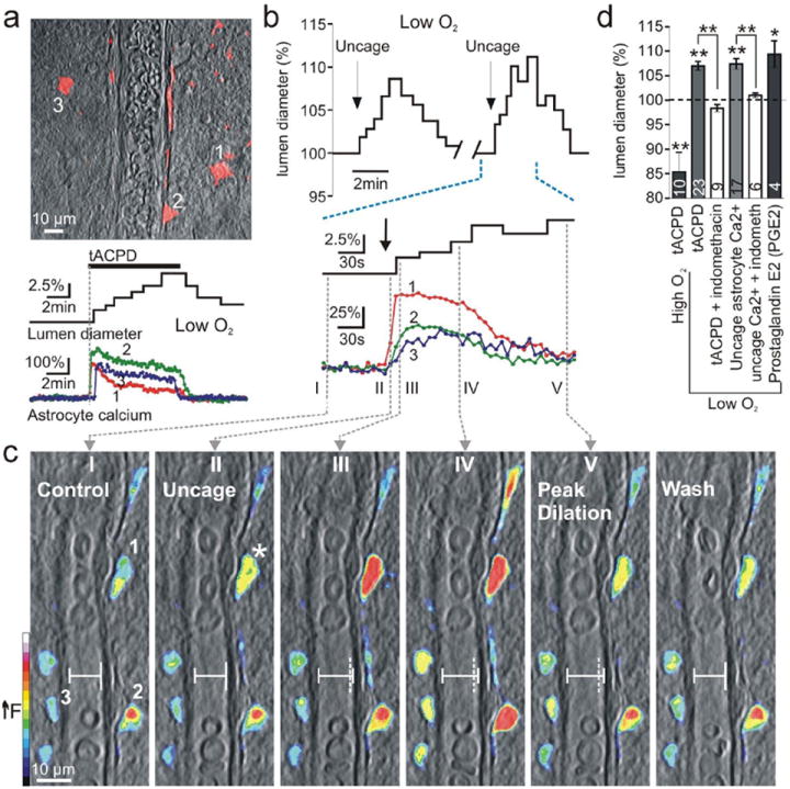
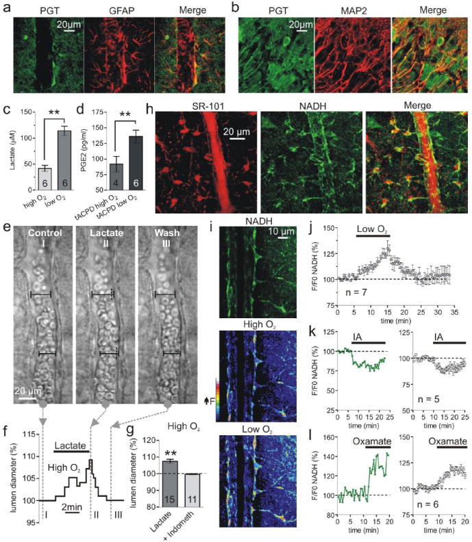
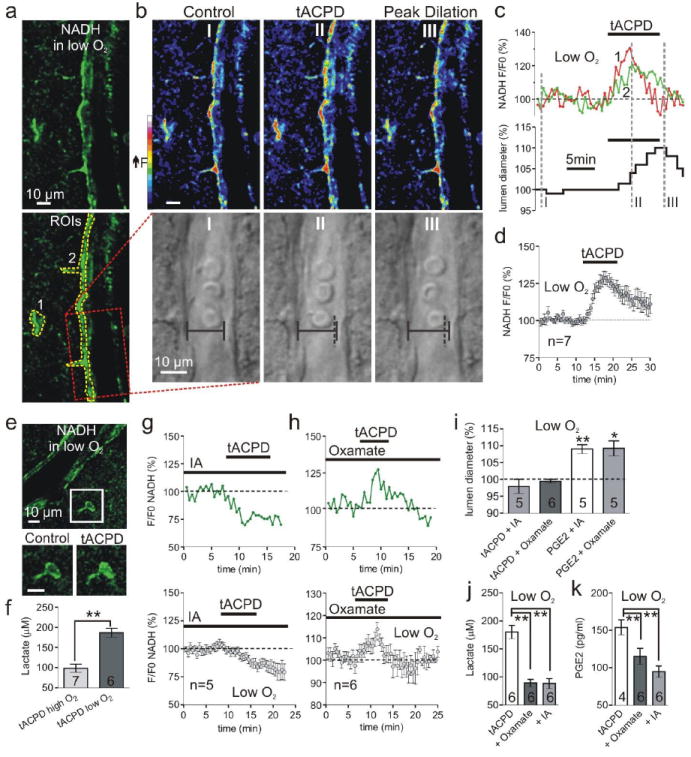
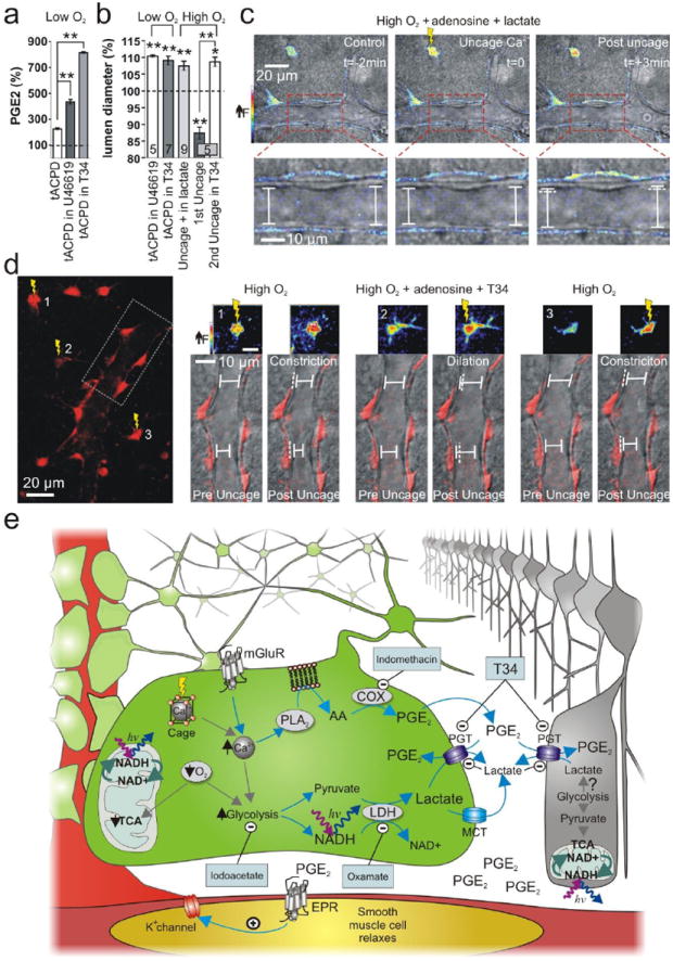
Calcium signalling in astrocytes couples changes in neural activity to alterations in cerebral blood flow by eliciting vasoconstriction or vasodilation of arterioles. However, the mechanism for how these opposite astrocyte influences provide appropriate changes in vessel tone within an environment that has dynamic metabolic requirements remains unclear. Here we show that the ability of astrocytes to induce vasodilations over vasoconstrictions relies on the metabolic state of the rat brain tissue. When oxygen availability is lowered and astrocyte calcium concentration is elevated, astrocyte glycolysis and lactate release are maximized. External lactate attenuates transporter-mediated uptake from the extracellular space of prostaglandin E(2), leading to accumulation and subsequent vasodilation. In conditions of low oxygen concentration extracellular adenosine also increases, which blocks astrocyte-mediated constriction, facilitating dilation. These data reveal the role of metabolic substrates in regulating brain blood flow and provide a mechanism for differential astrocyte control over cerebrovascular diameter during different states of brain activation.


Figures

Fig. 1
Fig. 1
Lowering O2 converts astrocyte-mediated vasoconstrictions to vasodilations. (a) Top: astrocytes (red) loaded with Ca2+ indicator surround arteriole. Bottom: astrocyte Ca2+ signals occur coincident with dilation caused by the mGluR agonist t-ACPD in low O2. (b) Top: uncaging astrocyte Ca2+ (indicated by arrow) causes vasodilation in low O2 and is repeatable. Bottom: expanded time scale shows the Ca2+ signal in endfoot 1 (shown in c I) precedes the lumen diameter increase. (c) Overlay of vessel and pseudo colored endfoot Ca2+ changes. Images I-V correspond to times in b. I: control state and regions of interest: endfeet 1-3. Image II: endfoot 1 shows first Ca2+ rise (star) before lumen diameter starts to increase. Vertical dotted line (III-V) indicates previous position of vessel wall. (d) Summary data.
Fig. 2
Fig. 2
Low O2 facilitates lactate and PGE2 release and enhances astrocyte glycolysis. (a and b) Immunohistochemistry showing the astrocyte marker GFAP and the neuron marker MAP2 colocalize with PGT labeling. (c) Lactate release is elevated in low O2. (d) t-APCD increases PGE2 release most in low O2. (e and f) Lactate dilates an arteriole. Images I-III correspond to time points in f, which shows the lumen diameter increase. (g) Summary showing percent dilation by lactate and block by indomethacin. (h) Colocalization of the astrocyte maker SR-101 (left stack) and the NADH signal (middle stack) from perivascular glia somas and endfeet. (i) Top: arteriole NADH fluorescence from a single image plane showing astrocyte compartments, SMCs/endothelial cells and empty lumen of the blood vessel. Middle and Lower: pseudo colored image of NADH fluorescence in high and low O2. (j) Summary of NADH changes in astrocyte compartments caused by low O2. (k and l) Astrocyte NADH in response to glycolysis inhibition with iodoacetate (IA) (k) and LDH inhibition with Oxamate (l). Left: single experiment; right: summary.
Fig. 3
Fig. 3
Glycolysis and lactate release is critical for astrocyte-mediated dilations. (a) Arteriole and astrocyte NADH image in low O2; bottom shows ROIs for astrocyte compartments (1 and 2) and vessel (box). (b) Pseudo color NADH (top) and diameter (bottom) changes caused by t-ACPD at time points I-III in c. Vertical dotted line (II and III) indicates previous position of vessel wall. (c) Astrocyte NADH (top) and lumen diameter (bottom) in response to t-ACPD; same experiment as a and b. (d) Summary of NADH increase from mGluR activation in low O2. (e) Top: NADH image of an arteriole and astrocyte. Lower: soma close-up showing t-ACPD causes a diffuse increase in NADH. (f) t-ACPD increases extracellular lactate most in low O2. (g) t-ACPD decreases astrocyte NADH during glycolysis inhibition; top: single experiment; bottom: summary. (h) mGluR activation increases astrocyte NADH during LDH inhibition; top: single experiment; bottom: summary. (i) Summary showing t-ACPD fails to dilate vessels in oxamate or IA and PGE2 rescues vasodilation in these compounds. (j and k) The increase in lactate (j) and PGE2 (k) caused by t-ACPD is significantly less in oxamate and IA.
Fig. 4
Fig. 4
Raising PGE2 levels by inhibiting PGT changes the polarity of astrocyte-mediating vasomotion. (a) PGE2 levels were further elevated by mGluR activation in low O2 when PGT was inhibited by U46619 or T34. (b) Summary data of vasomotion during PGT manipulation. (c) Uncaging Ca2+ in high O2 causes vasodilation in exogenous lactate. Top panels show the astrocyte Ca2+ signal change (pseudo colour) from uncaging. Lower panels show close up of vessel lumen. (d) In high O2, astrocyte-mediated vasoconstriction is converted to vasodilation during PGT blockade. Left: astrocytes and endfeet circumscribing an arteriole. Ca2+ is uncaged in 3 astrocytes separated in time; box indicates the vessel region examined on the right. Right: vasomotions corresponding to the separate uncaging events. Small pseudo colour images show the Ca2+ signal change from uncaging in each astrocyte. Lower images of the vessel and endfeet (red) show that the vasomotion switches polarity when PGTs are blocked. (e) Diagram of the supported model.

Comment in

  • Neuroscience: Brain power.
    Hall CN, Attwell D. Hall CN, et al. Nature. 2008 Dec 11;456(7223):715-6. doi: 10.1038/456715a. Nature. 2008. PMID: 19079043 No abstract available.

References

    1. Mulligan SJ, MacVicar BA. Calcium transients in astrocyte endfeet cause cerebrovascular constrictions. Nature. 2004;431(7005):195. - PubMed
    1. Metea MR, Newman EA. Glial cells dilate and constrict blood vessels: a mechanism of neurovascular coupling. J Neurosci. 2006;26(11):2862. - PMC - PubMed
    1. Chuquet J, Hollender L, Nimchinsky EA. High-resolution in vivo imaging of the neurovascular unit during spreading depression. J Neurosci. 2007;27(15):4036. - PMC - PubMed
    1. Zonta M, Angulo MC, Gobbo S, et al. Neuron-to-astrocyte signaling is central to the dynamic control of brain microcirculation. Nature neuroscience. 2003;6(1):43. - PubMed
    1. Filosa JA, Bonev AD, Nelson MT. Calcium dynamics in cortical astrocytes and arterioles during neurovascular coupling. Circulation research. 2004;95(10):e73. - PubMed

Publication types

MeSH terms