. 2008 Nov 19;27(22):2988-97.
doi: 10.1038/emboj.2008.230. Epub 2008 Oct 30.

Low reduction potential of Ero1alpha regulatory disulphides ensures tight control of substrate oxidation

Affiliations

Low reduction potential of Ero1alpha regulatory disulphides ensures tight control of substrate oxidation

Karl M Baker et al. EMBO J. .

Abstract

Formation of disulphide bonds within the mammalian endoplasmic reticulum (ER) requires the combined activities of Ero1alpha and protein disulphide isomerase (PDI). As Ero1alpha produces hydrogen peroxide during oxidation, regulation of its activity is critical in preventing ER-generated oxidative stress. Here, we have expressed and purified recombinant human Ero1alpha and shown that it has activity towards thioredoxin and PDI. The activity towards PDI required the inclusion of glutathione to ensure sustained oxidation. By carrying out site-directed mutagenesis of cysteine residues, we show that Ero1alpha is regulated by non-catalytic disulphides. The midpoint reduction potential (E degrees') of the regulatory disulphides was calculated to be approximately -275 mV making them stable in the redox conditions prevalent in the ER. The stable regulatory disulphides were only partially reduced by PDI (E degrees' approximately -180 mV), suggesting either that this is a mechanism for preventing excessive Ero1alpha activity and oxidation of PDI or that additional factors are required for Ero1alpha activation within the mammalian ER.


Figures

Figure 1
Figure 1
The redox state of Ero1α changes transiently during the efficient oxidation of thioredoxin. (A) Ero1α oxidises Trx1 (100 μM) as confirmed by the change of redox status judged by alkylation with AMS when incubated in the presence of Ero1α (0.5 μM) but not in its absence. Proteins were visualised by reducing SDS–PAGE and Coomassie blue staining. (B) Ero1α is transiently reduced following incubation with Trx1. Reactions were quenched with NEM, the redox state of Ero1α was visualised by non-reducing SDS–PAGE and silver staining. The reduced and oxidised forms of thioredoxin and Ero1α are indicated as r and ox, respectively. (A, B) Representative gels; the experiment was repeated at least three times with similar results.
Figure 2
Figure 2
Ero1α oxidises PDI and shows selectivity towards the a′ active site. (A) Kinetics of oxygen consumption by Ero1α (2 μM) during reaction with reduced PDI (100 μM) was assayed either in the absence (A) or presence (B) of GSH (10 mM). Control reactions following oxygen consumption in the absence of Ero1α are as indicated. (C) The oxygen consumption by Ero1α (2 μM) during reaction with either a ΔS1 or ΔS2 mutant of PDI in the presence of 10 mM GSH. The experiments were carried out three times with similar activity profiles.
Figure 3
Figure 3
Ero1α oxidises PDI but the long-range disulphides in Ero1α are only partially reduced. (A) The oxidation of PDI by Ero1α was followed by the change in redox state of reduced PDI as judged by the access of free thiols to alkylation by mPEG-mal. Reduced PDI (100 μM) was incubated in the absence (lanes 2 and 3) or presence (lanes 4–7) of Ero1α and in the absence (lanes 2–5) or presence (lanes 6 and 7) of GSH (10 mM). Reactions proceeded for 5 or 60 min as indicated before being terminated by the addition of TCA followed by alkylation with mPEG-mal. Protein was separated by reducing SDS–PAGE and visualised by Coomassie staining. An untreated and mPEG-mal-treated reduced PDI sample (lanes 1 and 2) and an mPEG-mal-treated sample of reduced ΔS1 and ΔS2 mutants of PDI (lanes 8 and 9) are shown to indicate the mobility shift on mPEG-mal modification of reduced PDI. (B) The redox state of Ero1α was assayed during the reaction with PDI either in the absence (lanes 1–4) or presence (lanes 5–7) of GSH (10 mM). Reactions were quenched with NEM and the redox state of Ero1α was visualised by non-reducing SDS–PAGE followed by western blotting with an anti-Ero1α antibody. Thioredoxin-reduced Ero1α was loaded in lane 8 for comparison. (C) The redox state of Ero1α was determined after incubation (15 min) with increasing concentrations of GSH (titrated to pH 7.5) as indicated. Reactions were quenched with NEM and the redox state of Ero1α was visualised by non-reducing SDS–PAGE and silver staining. The experiments in (A–C) were repeated three times with similar results obtained.
Figure 4
Figure 4
Identification of the cysteine residues involved in the formation of long-range disulphides in Ero1α. (A) Reduced (lanes 10 and 14) and non-reduced (lanes 1 and 13) wild-type Ero1α were separated by SDS–PAGE along with non-reduced cysteine mutants of Ero1α as indicated. Protein was visualised following SDS–PAGE and Coomassie staining. (B) Schematic illustration comparing the disulphide bonds within human Ero1α and yeast Ero1p that are either active site disulphides (formula image) or regulatory disulphides (formula image
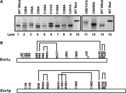
). Known disulphide pairings are as indicated as they exist in the inactive state of the protein. Note that in the human protein cys94 and cys99 are engaged in disulphide pairings with cys131 and cys104, respectively, whereas in the active state these would form a disulphide with each other.
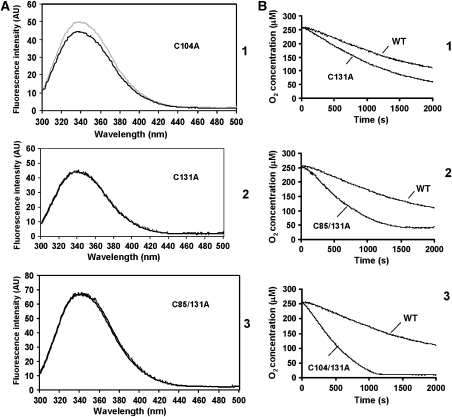
Figure 5
Figure 5
Ero1α long-range disulphides regulate conformation changes and enzymatic activity. (A) The fluorescence emission spectra of reduced (grey line) or non-reduced (black line) Ero1α mutants C104A (1), C131A (2) and C85/131A (3) were analysed following excitation at 280 nm. (B) Oxygen consumption was assayed with PDI (100 μM) in the presence of 10 mM GSH and the Ero1α mutants (2 μM) C131A (1), C85/131A (2) or C104/131 (3). These are representative profiles from three separate experiments. The activity of wild-type Ero1α towards PDI is also included in each graph as indicated.
Figure 6
Figure 6
Measuring the midpoint reduction potential of the regulatory disulphides in Ero1α. (A) The enzymatically inactive C99A mutant (1 μM) was equilibrated with various ratios of reduced/oxidised thioredoxin before alkylation with NEM and separation by non-reducing SDS–PAGE. Protein was visualised by Coomassie staining and the fraction of reduced Ero1α was quantified by densitometry. (B) Values for the fraction of reduced Ero1α were plotted against the ratio of reduced/oxidised thioredoxin. Plots were interpreted by equation (1), and Keq values were determined as described. The experiment was repeated three times and the average Keq was used to calculate the reduction potential.

Comment in

References

    1. Appenzeller-Herzog C, Ellgaard L (2008) In vivo reduction–oxidation state of protein disulphide isomerase: the two active sites independently occur in the reduced and oxidized forms. Antioxid Redox Signal 10: 55–64 - PubMed
    1. Appenzeller-Herzog C, Riemer J, Christensen B, Sørensen ES, Ellgaard L (2008) A novel disulphide switch mechanism in Ero1α balances ER oxidation in human cells. EMBO J (advance online publication, 2 October 2008; doi:10.1038/emboj.2008.202) - PMC - PubMed
    1. Benham AM, Cabibbo A, Fassio A, Bulleid N, Sitia R, Braakman I (2000) The CXXCXXC motif determines the folding, structure and stability of human Ero1-Lalpha. EMBO J 19: 4493–4502 - PMC - PubMed
    1. Bertoli G, Simmen T, Anelli T, Molteni SN, Fesce R, Sitia R (2004) Two conserved cysteine triads in human Ero1alpha cooperate for efficient disulphide bond formation in the endoplasmic reticulum. J Biol Chem 279: 30047–30052 - PubMed
    1. Bradford MM (1976) A rapid and sensitive method for the quantitation of microgram quantities of protein utilizing the principle of protein–dye binding. Anal Biochem 72: 248–254 - PubMed

Publication types

MeSH terms