. 2008 Dec 3;27(23):3129-39.
doi: 10.1038/emboj.2008.228. Epub 2008 Oct 30.

A protein that replaces the entire cellular eIF4F complex

Affiliations

A protein that replaces the entire cellular eIF4F complex

Mohammad A Mir et al. EMBO J. .

Abstract

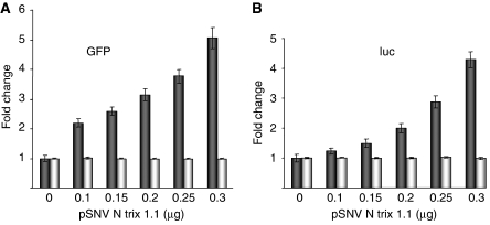
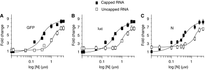
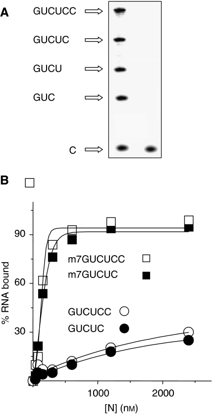
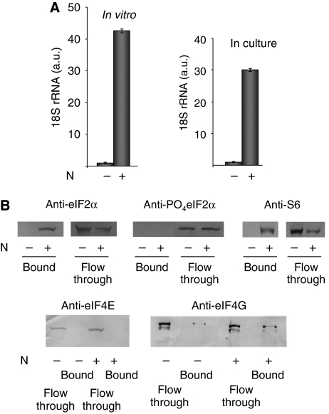
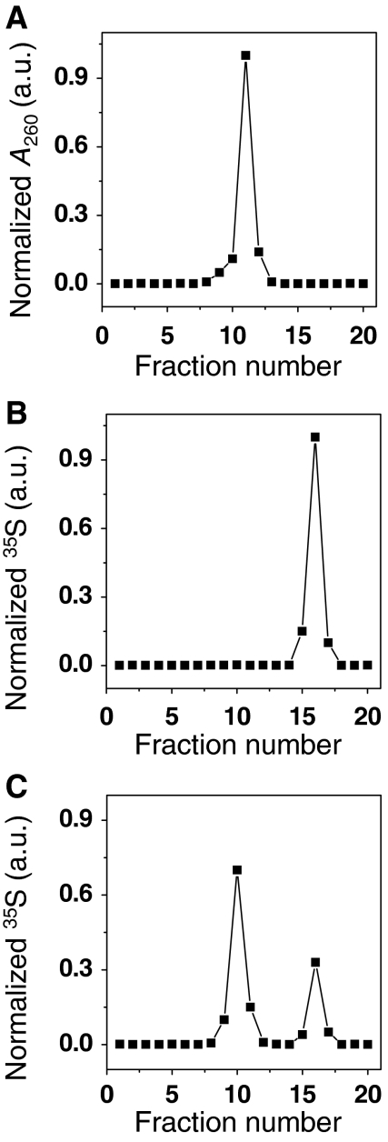
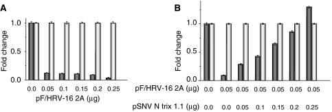
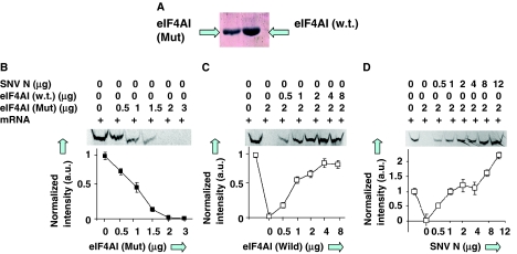
The eIF4F cap-binding complex mediates the initiation of cellular mRNA translation. eIF4F is composed of eIF4E, which binds to the mRNA cap, eIF4G, which indirectly links the mRNA cap with the 43S pre-initiation complex, and eIF4A, which is a helicase necessary for initiation. Viral nucleocapsid proteins (N) function in both genome replication and RNA encapsidation. Surprisingly, we find that hantavirus N has multiple intrinsic activities that mimic and substitute for each of the three peptides of the cap-binding complex thereby enhancing the translation of viral mRNA. N binds with high affinity to the mRNA cap replacing eIF4E. N binds directly to the 43S pre-initiation complex facilitating loading of ribosomes onto capped mRNA functionally replacing eIF4G. Finally, N obviates the requirement for the helicase, eIF4A. The expression of a multifaceted viral protein that functionally supplants the cellular cap-binding complex is a unique strategy for viral mRNA translation initiation. The ability of N to directly mediate translation initiation would ensure the efficient translation of viral mRNA.


Figures

Figure 1
Figure 1
N increases the expression of reporter proteins. HeLa cells were transfected with a constant amount of reporter plasmid and increasing amounts of a plasmid expressing hantavirus N. Evaluation of N expression on western blots with anti-N antibody indicated that N expression increased along with increasing amounts of plasmid, as expected (not shown). At 36 h after transfection, cells were harvested and GFP or luc expression was quantified, by flow cytometry or enzymatically, respectively (dark bars). (A) Expression of GFP is shown. (B) luc as a function of increasing N is shown. Steady-state GFP mRNA and luc mRNA were quantified using ‘real-time' RT–PCR with primers specific for a segment in the centre of each reporter RNA. In both (A, B), the results of this latter analysis are depicted with light bars.
Figure 2
Figure 2
N augments the translational expression of capped mRNA. We examined the effect of increasing N on the translational expression of three reporter mRNAs containing or lacking a 5′ cap using rabbit reticulocyte lysates. Capped and uncapped mRNA encoding GFP, luc or N were translated in vitro in the presence of 35S-methionine and increasing amounts of N in (AC), respectively. Translation products were then electrophoresed on SDS polyacrylamide gels, and the amount of translation product was quantified by phosphorimage analysis. Translation of capped and uncapped RNA is depicted with filled and open squares, respectively. The amount of labelled protein synthesized with capped RNA in reactions lacking N was normalized to 1 (this cannot be indicated on the log scale). In the absence of N, expression of the indicator proteins was slightly higher with uncapped than capped RNA. This is consistent with earlier observations (Svitkin et al, 1996). Thus, the amount of expression at the lower concentrations of N is equivalent to background levels of expression from capped and uncapped RNA for each of the indicator RNAs.
Figure 3
Figure 3
N binds to capped oligoribonucleotides. (A) Short radioactively labelled capped and uncapped RNAs were synthesized using T7 RNA polymerase and α-32P-CTP. As the third nucleotide of transcription products arising from the T7 promoter is the first C residue of the RNA, only molecules 3 nt long or greater were labelled. These short RNAs were separated on, and recovered from, a high percentage denaturing polyacrylamide gel. The image depicts such a gel. The leftward lane displays the series of short oligoribonucleotides arising from transcription. The rightward lane contains unincorporated CTP as a migration control. (B) Each RNA was incubated with increasing concentrations of purified N, and association of RNA with N was quantified by filter binding. Binding of N with capped (m7GUCUCC) or uncapped (GUCUCC) are indicated with open squares and circles, respectively, and with m7GUCUC and GUCUC with closed squares and circles, respectively. Binding experiments carried out with both capped and uncapped RNAs less than 5 nt in length exhibited negligible binding with N. For example, binding with the 4-nt long RNA, GUCU, in capped and uncapped form is shown in Supplementary Figure S1.
Figure 4
Figure 4
N preferentially augments the translation of viral mRNA. (A) Equimolar amounts of capped mRNA containing the 5′ untranslated region from S segment mRNA and encoding N (v-N), and an mRNA containing a non-viral leader region and encoding GFP (GFP) were added together to reticulocyte extracts containing increasing concentrations of N as indicated. The concentration of each mRNA was approximately 45 nM. Labelled N and GFP were separated by PAGE and quantified by phosphorimage analysis (shown below the graph). Similar results were obtained in three separate experiments. In (B), the leader regions from the mRNAs of (A) were interchanged. Thus, one mRNA contained the 5′ viral UTR preceding the GFP gene (v-GFP), and a second mRNA contained the non-viral leader preceding the N gene (N). Translation and quantitation were as in (A). (C) Radioactively labelled capped RNAs were used in binding reactions with purified N and the binding affinity (Kd) was determined for each. Viral sequences are shown schematically in grey, whereas non-viral sequences are in white. n-v-GFP contains a 9 nt non-viral cap simulating cellular RNA derived from cap-snatching (shown in black). Note: The leader regions are not shown to scale relative to the N and GFP genes. The untranslated leaders, GFP gene, and N gene are 43, 798, and 1287 nt in length, respectively. (D) Comparison of the leader sequences from GFP, v-GFP, and n-v-GFP mRNA and minus strand S segment viral RNA. Nucleotides required for high-affinity binding to the vRNA panhandle are depicted with shading and include nucleotides from both the 5′ and 3′ termini (Mir and Panganiban, 2005). The 5′ terminal nucleotides of +strand mRNA required for binding by N is also indicated by shading. As the termini of the viral genome segments consist of imperfect inverted repeats, the 5′ sequences of both plus and minus strand viral RNA are similar. Nucleotide differences in the 5′ sequence of mRNA relative to the 5′ sequence of minus strand vRNA are indicated with bold lettering. Leader sequences of v-GFP and n-v-GFP. The 9-nt-long non-viral leader of n-v-GFP, and the start codon of the mRNAs are underlined.
Figure 5
Figure 5
N interacts with the pre-initiation complex. (A) N was incubated with rabbit reticulocyte lysates and recovered with Ni-NTA beads. The bound material was eluted from the Ni-NTA, RNA was purified, and 18S rRNA was quantified using real-time RT–PCR. The leftward graph depicts the relative amount of 18S rRNA associated with Ni-NTA beads in the absence and presence of N. The rightward graph depicts an analogous experiment carried out with 293 cells that were transfected with either an N-expressing plasmid or its parental vector, as a negative control. N was recovered from the lysates of transfected cells using Ni-NTA and 18S rRNA that co-purified with N quantified by real-time RT–PCR. (B) A set of western blots to examine the association of peptide constituents of the 43S pre-initiation complex, and the eIF4F cap-binding complex, that co-purify with N. N was expressed by transfection, isolated from the lysates of these cells using Ni-NTA columns, bound material was recovered and subjected to western blot analysis with primary antibodies as indicated. Peptides that co-purify with N (bound), or that flow through the column are indicated.
Figure 6
Figure 6
N binds directly to the small ribosomal subunit. 40S small ribosomal subunits were prepared by incubation of ribosomes in the presence of puromycin and purified from large ribosomal subunits and mRNA. (A) Purified 40S subunits were then resedimented on a sucrose gradient. (B) N protein was expressed by in vitro translation in the presence of 35S-methionine, purified from the translation mixture by denaturation with urea, recovery on Ni-NTA beads, renaturation by dialysis and sedimented in parallel with 40S subunits. (C) N was incubated with excess purified 40S subunits prior to sedimentation. Leftward fractions correspond to those from the bottom of the gradient.
Figure 7
Figure 7
N replaces eIF4G. (A) HeLa cells were co-transfected with a plasmid expressing reporter GFP, along with increasing amounts of pF/HRV-16 2A, which expresses the 2A protease of HRV-16. GFP expression was quantified using flow cytometry as in Figure 1 (dark bars) and GFP mRNA was quantified using real-time PCR (light bars). (B) Cells were transfected with a constant amount of GFP expression plasmid, a constant amount of 2A expression plasmid (0.05 μg) sufficient for significantly reducing translation of the reporter gene, and increasing amounts of an N expression plasmid. GFP expression (dark bars) and steady-state GFP mRNA (light bars) were quantified as in (A). In the experiments of (A, B), the total amount of DNA used in the transfections was held constant by the addition of parental vector.
Figure 8
Figure 8
N promotes ribosome loading. A synthetic mRNA containing 3′ poly A was incubated in reticulocyte lysates to allow translation. The synthetic polyadenylated RNA was recovered from the translation mixture using oligo dT beads. Ribosomes associated with the polyadenylated RNA were quantified by real-time RT–PCR with primer sets specific for 18 and 28S rRNA.
Figure 9
Figure 9
N functionally replaces eIF4A. (A) Bacterially expressed and purified wild-type and mutant eIF4A were used in in vitro translation reactions containing luciferase mRNA. (B) Effect of dominant-negative mutant eIF4A on translation. Translation was quantified by SDS–PAGE followed by phosphorimage analysis of radioactively labelled luc. (C) Similar reactions were carried out in the presence of fixed amount (2 μg) of mutant eIF4A and increasing amounts of wild-type eIF4A. (D) Translation reactions in the presence of 2 μg of mutant eIF-4A and increasing amounts of N.

References

    1. Ali IK, McKendrick L, Morley SJ, Jackson RJ (2001) Truncated initiation factor eIF4G lacking an eIF4E binding site can support capped mRNA translation. EMBO J 20: 4233–4242 - PMC - PubMed
    1. Barr JN (2007) Bunyavirus mRNA synthesis is coupled to translation to prevent premature transcription termination. RNA 13: 731–736 - PMC - PubMed
    1. Bellocq C, Kolakofsky D (1987) Translational requirement for La Crosse virus S-mRNA synthesis: a possible mechanism. J Virol 61: 3960–3967 - PMC - PubMed
    1. Blakqori G, Kochs G, Haller O, Weber F (2003) Functional L polymerase of La Crosse virus allows in vivo reconstitution of recombinant nucleocapsids. J Gen Virol 84 (Part 5): 1207–1214 - PubMed
    1. Bochkov YA, Palmenberg AC (2006) Translational efficiency of EMCV IRES in bicistronic vectors is dependent upon IRES sequence and gene location. Biotechniques 41: 283–284, 286, 288 passim - PubMed

Publication types

MeSH terms

Substances