

Review
doi: 10.1007/s00441-008-0706-5.
Epub 2008 Oct 30.
Arterial versus venous endothelial cells
Affiliations
- PMID: 18972135
- PMCID: PMC4105978
- DOI: 10.1007/s00441-008-0706-5
Item in Clipboard
Review
Arterial versus venous endothelial cells
Cell Tissue Res.
2009 Jan.
Abstract
Vascular endothelial cells (ECs) form the inner lining of all blood vessels from the largest artery and veins, viz., the aorta and venae cavae, respectively, to the capillaries that connect the arterial and venous systems. Because these two major conducting systems of the cardiovasculature differ functionally, it is not surprising that the physical makeup of arteries and veins, including the ECs that line their lumina, are also distinct. Although few would argue that the local environment contributes to the differences between arteries and veins, recent evidence has shown that the specification of arterial and venous identity is largely genetically determined.
Figures
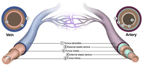
Structural features of the vasculature. All blood vessels within the branches of the vascular tree, except the capillaries, are composed of a tunica intima that is lined with endothelial cells (ECs), a tunica media that contains SMCs and elastic fibers, and a tunica adventitia made up of fibrous connective tissue. Arterial blood vessels (dark red/purple) are characterized by long narrow ECs that are aligned in the direction of blood flow, multiple layers of smooth muscle cells, and in the case of muscular arteries, elastic fibers that are arranged in two distinct bands (inner and outer elastic laminae). In contrast, venous blood vessels (blue) are lined with rounder non-aligned ECs, lack elastic laminae, and most possess valves that project into their lumen

Endomucin is selectively expressed by veins but not arteries. Diaminobenzidine immunohistochemical staining shows endomucin expression in a mouse pancreatic vein (arrow). Expression in the adjacent artery (arrowhead) is absent. Scale bar, 50 μm

Model of arteriovenous EC specification during embryonic development. Angioblasts derived from the lateral plate mesoderm (LPM) are specified into either arterial or venous ECs prior to migration to the midline where they form the dorsal aorta (DA) and posterior cardinal vein (PCV). Specification is initiated by the expression of sonic hedgehog (Shh) by the notochord (Nc) and floor plate of the neural tube (NT), with Shh then acting on the adjacent somites (S) to induce the expression of vascular endothelial growth factor (VEGF). In arterial-fated angioblasts, VEGF signaling via a VEGF receptor 2 (VEGFR2)/neuropilin 1 (NP-1) complex leads to the downstream activation of Notch and ERK signaling and the subsequent expression of the arterial marker ephrinB2. Foxc1 and Foxc2 (Foxc1/2) are transcriptional factors that control the expression of Notch components and therefore also promote arterial EC identity. In venous-fated angioblasts, COUP-TFII blocks NP-1 expression, which prevents Notch and ERK activation, and the venous marker EphB4 is expressed. Activation of the PI3K/AKT pathway also blocks ERK, which represses arterial EC fate. Adapted from Lamont and Childs (2006) and Lawson and Weinstein (2002)
References
-
- Ahn DG, Ruvinsky I, Oates AC, Silver LM, Ho RK. tbx20, a new vertebrate T-box gene expressed in the cranial motor neurons and developing cardiovascular structures in zebrafish. Mech Dev. 2000;95:253–258. - PubMed
-
- Aird WC. Phenotypic heterogeneity of the endothelium. I. Structure, function, and mechanisms. Circ Res. 2007;100:158–173. - PubMed
-
- Alon T, Hemo I, Itin A, Pe’er J, Stone J, Keshet E. Vascular endothelial growth factor acts as a survival factor for newly formed retinal vessels and has implications for retinopathy of prematurity. Nat Med. 1995;1:1024–1028. - PubMed
-
- Artavanis-Tsakonas S, Rand MD, Lake RJ. Notch signaling: cell fate control and signal integration in development. Science. 1999;284:770–776. - PubMed
Publication types
MeSH terms
Grants and funding
LinkOut - more resources
Full Text Sources
Other Literature Sources
