. 2008 Nov;28(5):1266-72.
doi: 10.1002/jmri.21576.

High-resolution 7T MRI of the human hippocampus in vivo

Affiliations

High-resolution 7T MRI of the human hippocampus in vivo

Bradley P Thomas et al. J Magn Reson Imaging. 2008 Nov.

Abstract

Purpose: To describe an initial experience imaging the human hippocampus in vivo using a 7T magnetic resonance (MR) scanner and a protocol developed for very high field neuroimaging.

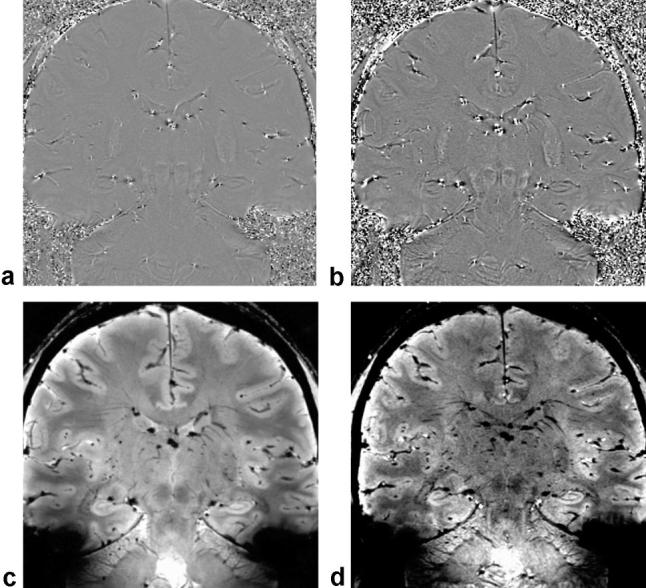
Materials and methods: Six normal subjects were scanned on a 7T whole body MR scanner equipped with a 16-channel head coil. Sequences included a full field of view T1-weighted 3D turbo field echo (T1W 3D TFE: time of acquisition (TA)=08:58), T2*-weighted 2D fast field echo (T2*W 2D FFE: TA=05:20), and susceptibility-weighted imaging (SWI: TA=04:20). SWI data were postprocessed using a minimum intensity projection (minIP) algorithm. Total imaging time was 23 minutes.

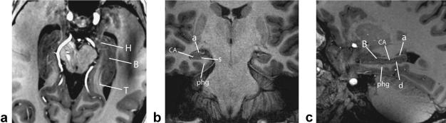
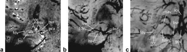
Results: T1W 3D TFE images with 700 microm isotropic voxels provided excellent anatomic depiction of macroscopic hippocampal structures. T2*W 2D FFE images with 0.5 mm in-plane resolution and 2.5 mm slice thickness provided clear discrimination of the Cornu Ammonis and the compilation of adjacent sublayers of the hippocampus. SWI images (0.5 mm in-plane resolution, 1.0 mm slice thickness) delineated microvenous anatomy of the hippocampus.

Conclusion: In vivo 7T MR imaging can take advantage of higher signal-to-noise and novel contrast mechanisms to provide increased conspicuity of hippocampal anatomy.


Figures

Figure 1
Figure 1
Hippocampal body, gross (left) and schematic (right) coronal-oblique views showing Cornu Ammonis (CA), subfields CA1–4 (–4), alveus (al), dentate gyrus (d), fimbria (f), fimbriodentate sulcus (fds), hippocampal sulcus (hs), parahippocampal gyrus (phg), basal vein of Rosenthal (bvr), stratum pyramidale (sp), compilation of strata radiatum, lacunosum, and moleculare (srlm), vein of temporal horn (vth), margo denticulatus (md), vein of superficial hippocampal sulcus (vshs), vein of fimbriodentate sulcus (vfds), medial atrial vein (mav), posterior hippocampal artery (pha), middle hippocampal artery (mha), inferior ventricular vein (ivv), posterior cerebral artery (pca), basilar artery (ba), entorhinal area (eh), superficial medullary lamina (sml), subiculum (s).
Figure 2
Figure 2
Left: Coronal H&E (Hemotoxin and Eosin) stained sections through the hippocampal body with enlargement of boxed area (right) showing alveus (1), stratum oriens (2), stratum pyramidale (3), stratum radiatum (4), stratum lacunosum (5), stratum moleculare (6) of Cornu Ammonis, stratum moleculare (7) of dentate gyrus, stratum granulosum (8), polymorphic layer (9), sulcal intrahippocampal vein (10), subfields of Cornu Ammonis (CA1–CA4), fimbria (f), dentate gyrus (d), hilar region (h), subiculum (s), artifactual separations during tissue processing (*), vestigial hippocampal sulcus (arrowheads).
Figure 3
Figure 3
Axial (a), coronal (b), and sagittal (c) in vivo 7T T1W 3D TFE imaging through the entire hippocampus (left) and body (center and right) showing detailed structures including hippocampal head (H), body (B), tail (T), Cornu Ammonis (CA), alveus (a), subiculum (s), parahippocampal gyrus (phg), and dentate gyrus (d).
Figure 4
Figure 4
In vivo 7T T2*W 2D FFE (a,d), T1W 3D TFE (b,e) coronal imaging of hippocampal head (top row) and body (bottom row) with comparable slices through gross specimens (c,f) showing the temporal horn of lateral ventricle (th), Cornu Ammonis (CA), alveus (al), amygdala (a), internal digitations of hippocampal head (id), band of Giacomini (bg), uncus (u), uncal sulcus (us), external digitations of hippocampal head (ed), subiculum (s), dentate gyrus (d), composite of strata radiatum, lacunosum, moleculare, and vestigial hippocampal sulcus (srlms), fimbria (f), hippocampal sulcal vein (hsv), and parahippocampal gyrus (pg).
Figure 5
Figure 5
In vivo microvenous anatomy imaged coronally with 7T SWI (a) and reconstructed 20-mm thick minimum intensity projections (minIPs) of the hippocampal head (b) and body regions (c) showing the following venous structures: fimbriodentate vein (fdv), intrahippocampal sulcal vein (isv), inferior ventricular vein (ivv), hippocampal sulcal vein (hsv), basal vein of Rosenthal (bvr), posterior cerebral artery (pca), medial atrial vein (mav), great cerebral vein of Galen (gcv), internal cerebral vein (icv).
Figure 6
Figure 6
Highpass-filtered phase images from the 2D FFE (a) and 3D SWI (b) sequences. The highpass-filtering step eliminates slowly varying background phase from sources such as static field inhomogeneity. The phase images exhibit discernable gray-white contrast, and the hippocampus is clearly depicted. The srlms is visible as a dark band. The corresponding 2D FFE magnitude image appears in (c) along with the phase-enhanced SWI magnitude in (d). In the phase-enhanced SWI magnitude image the venous structures have slightly increased contrast with respect to background tissue.

References

    1. Abduljalil AM, Schmalbrock P, Novak V, Chakeres DW. Enhanced gray and white matter contrast of phase susceptibility-weighted images in ultra-high-field magnetic resonance imaging. J Magn Reson Imaging. 2003;18:284–290. - PubMed
    1. Novak V, Abduljalil AM, Novak P, Robitaille PM. High-resolution ultrahigh-field MRI of stroke. Magn Reson Imaging. 2005;23:539–548. - PMC - PubMed
    1. Wiesinger F, Van de Moortele PF, Adriany G, De Zanche N, Ugurbil K, Pruessmann KP. Parallel imaging performance as a function of field strength—an experimental investigation using electrodynamic scaling. Magn Reson Med. 2004;52:953–964. - PubMed
    1. Shaw CM, Alvord EC., Jr. Neuropathology of the limbic system. Neuroimaging Clin N Am. 1997;7:101–142. - PubMed
    1. Duvernoy HM. The human hippocampus: functional anatomy, vascularization and serial sections with MRI. 3rd ed. Springer; Heidelberg: 2005.

Publication types