

Therapeutic modulation of apoptosis: targeting the BCL-2 family at the interface of the mitochondrial membrane
- PMID: 18972587
- PMCID: PMC2615374
- DOI: 10.3349/ymj.2008.49.5.689
Therapeutic modulation of apoptosis: targeting the BCL-2 family at the interface of the mitochondrial membrane
Abstract
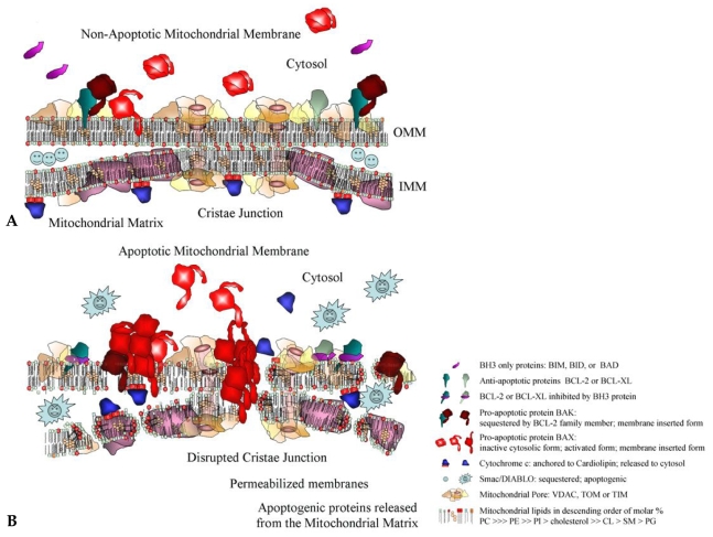
A vast portion of human disease results when the process of apoptosis is defective. Disorders resulting from inappropriate cell death range from autoimmune and neurodegenerative conditions to heart disease. Conversely, prevention of apoptosis is the hallmark of cancer and confounds the efficacy of cancer therapeutics. In the search for optimal targets that would enable the control of apoptosis, members of the BCL-2 family of anti- and pro-apoptotic factors have figured prominently. Development of BCL-2 antisense approaches, small molecules, and BH3 peptidomimetics has met with both success and failure. Success-because BCL-2 proteins play essential roles in apoptosis. Failure-because single targets for drug development have limited scope. By examining the activity of the BCL-2 proteins in relation to the mitochondrial landscape and drawing attention to the significant mitochondrial membrane alterations that ensue during apoptosis, we demonstrate the need for a broader based multi-disciplinary approach for the design of novel apoptosis-modulating compounds in the treatment of human disease.
Figures
References
-
- Metzstein MM, Stanfield GM, Horvitz HR. Genetics of programmed cell death in C. elegans: past, present and future. Trends Genet. 1998;14:410–416. - PubMed
-
- Yuan J, Shaham S, Ledoux S, Ellis HM, Horvitz HR. The C. elegans cell death gene ced-3 encodes a protein similar to mammalian interleukin-1 beta-converting enzyme. Cell. 1993;75:641–652. - PubMed
-
- Yuan J, Horvitz HR. The Caenorhabditis elegans cell death gene ced-4 encodes a novel protein and is expressed during the period of extensive programmed cell death. Development. 1992;116:309–320. - PubMed
-
- Hengartner MO, Horvitz HR. C. elegans cell survival gene ced-9 encodes a functional homolog of the mammalian proto-oncogene bcl-2. Cell. 1994;76:665–676. - PubMed
-
- Conradt B, Horvitz HR. The C. elegans protein EGL-1 is required for programmed cell death and interacts with the Bcl-2-like protein CED-9. Cell. 1998;93:519–529. - PubMed
Publication types
MeSH terms
Substances
Grants and funding
LinkOut - more resources
Full Text Sources
Miscellaneous
