

Dual simulated childbirth injuries result in slowed recovery of pudendal nerve and urethral function
- PMID: 18973146
- PMCID: PMC2661359
- DOI: 10.1002/nau.20632
Dual simulated childbirth injuries result in slowed recovery of pudendal nerve and urethral function
Abstract
Aims: Pelvic floor muscle trauma and pudendal nerve injury have been implicated in stress urinary incontinence (SUI) development after childbirth. In this study, we investigated how combinations of these injuries affect recovery.
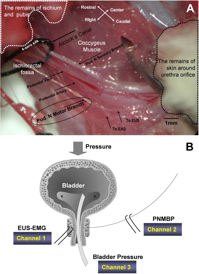
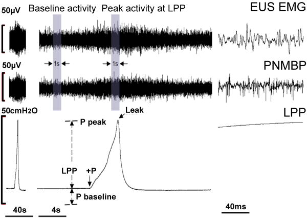
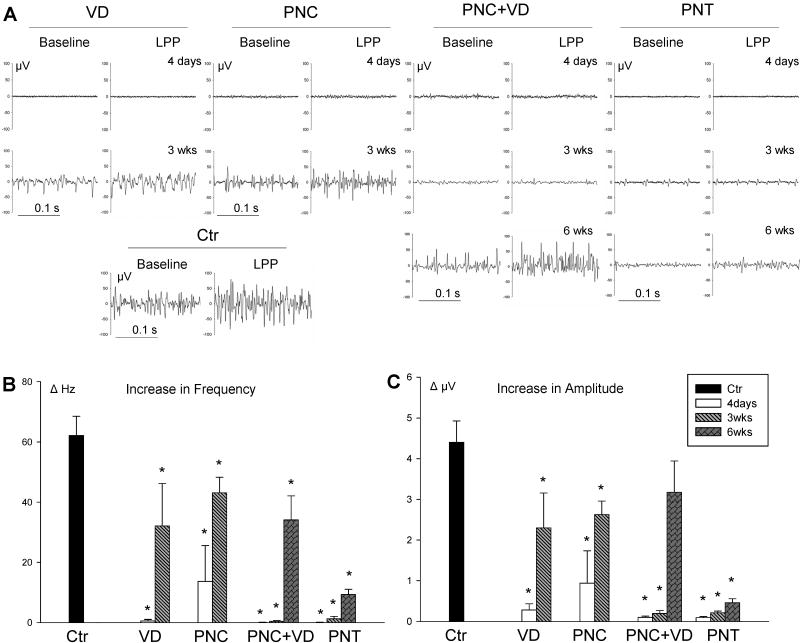
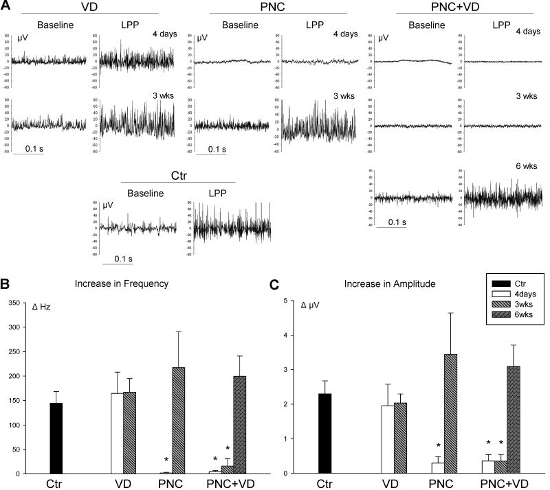
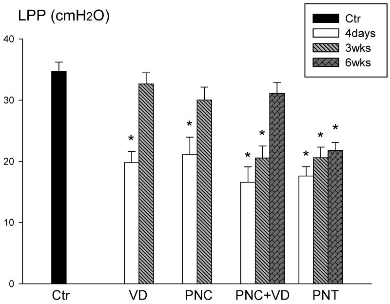
Methods: Sixty-seven female Sprague-Dawley rats underwent vaginal distension (VD), pudendal nerve crush (PNC), PNC and VD (PNC + VD), pudendal nerve transection (PNT), or served as unmanipulated controls. Four days, 3 weeks, or 6 weeks after injury, we simultaneously recorded pudendal nerve motor branch potentials (PNMBP), external urethral sphincter electromyography (EUS EMG), and transurethral bladder pressure under urethane anesthesia. The presence of a guarding reflex (increased frequency and amplitude of PNMBP or EUS EMG activity) during leak point pressure (LPP) testing was determined.
Results: Controls consistently demonstrated a guarding reflex. Four days after VD, EUS EMG activity was eliminated, but PNMBP activity reflected the guarding reflex; EUS EMG activity recovered after 3 weeks. Four days after PNC, both EUS EMG and PNMBP activity were eliminated, but demonstrated significant recovery at 3 weeks. Four days after PNC + VD both EUS EMG and nerve activity were eliminated, and little recovery was observed after 3 weeks with significant recovery of the guarding reflex 6 weeks after injury. Little recovery was observed at all time points after PNT. LPP results mirrored the reduction in EUS EMG activity.
Conclusion: Functional recovery occurs more slowly after PNC + VD than after either PNC or VD alone. Future work will be aimed at testing methods to facilitate neuroregeneration and recovery after this clinically relevant dual injury.
Figures
References
-
- Retzky SS, Rogers RMJ. Urinary Incontinence in Women. 47. Summit, NJ: Ciba-Geigy Corp.; 1995. - PubMed
-
- Damaser MS, Whitbeck C, Chichester P, Levin RM. Effect of vaginal distension on blood flow and hypoxia of urogenital organs of the female rat. J Appl Physiol. 2005 May;98(5):1884–90. - PubMed
-
- Lien KC, Morgan DM, DeLancey JO, Ashton-Miller JA. Pudendal nerve stretch during vaginal birth: a 3D computer simulation. American Journal of Obstetrics & Gynecology. 2005;192(5):1669–76. - PubMed
-
- Dietz HP, Simpson JM. Does delayed child-bearing increase the risk of levator injury in labour? Aust N Z J Obstet Gynaecol. 2007 Dec;47(6):491–5. - PubMed
-
- Snooks SJ, Swash M, Mathers SE, Henry MM. Effect of vaginal delivery on the pelvic floor: a 5-year follow-up. British Journal of Surgery. 1990;77:1358–60. - PubMed
Publication types
MeSH terms
Grants and funding
LinkOut - more resources
Full Text Sources
Other Literature Sources
Medical
