

Feasibility of myxomatous mitral valve repair using direct leaflet and chordal radiofrequency ablation
- PMID: 18973508
- PMCID: PMC2602795
- DOI: 10.1111/j.1540-8183.2008.00398.x
Feasibility of myxomatous mitral valve repair using direct leaflet and chordal radiofrequency ablation
Abstract
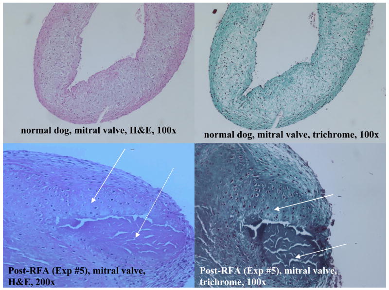
Objective: Minimally invasive repair of mitral valve prolapse (MVP) causing severe mitral regurgitation (MR) should reduce MR and have chronic durability. Our ex vivo, acute in vivo, and chronic in vivo studies suggest that direct application of radiofrequency ablation (RFA) to mitral leaflets and chordae can effect these repair goals to decrease MR.
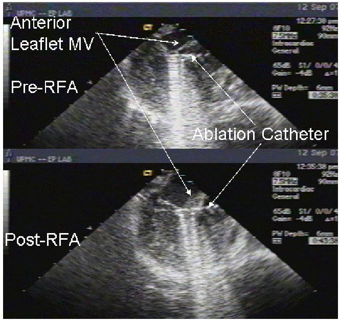
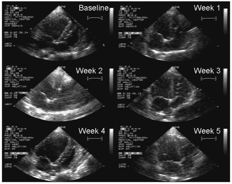
Methods: A total of seven canines were studied to assess the effects of RFA on mitral valve structure and function. RFA was applied ex vivo (n = 1), acutely in vivo using a right lateral thoracotomy and cardiopulmonary bypass (n = 3), and chronically in vivo using percutaneous access to the heart (n = 3). RFA was applied to the mitral valve and its associated chordae. Mitral valve structure and function (in vivo preparations) were then assessed.
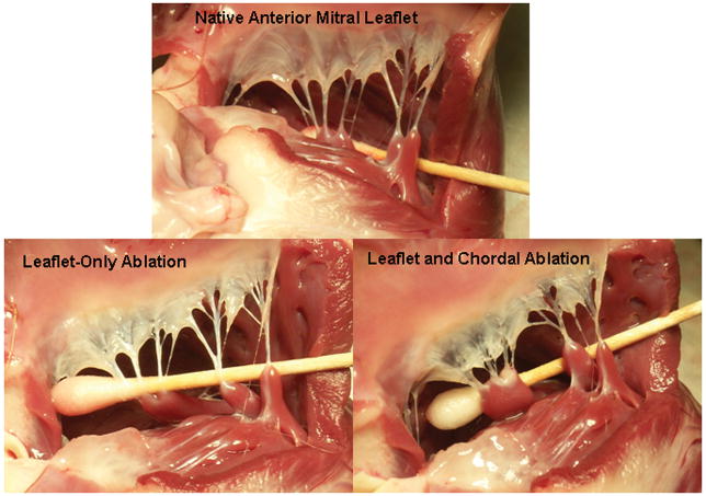
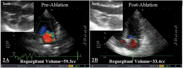
Results: Ex vivo application of RFA resulted in qualitative reduction in mitral leaflet surface area and chordal length. Acute in vivo application of RFA to canines found to have MVP causing severe MR demonstrated a 43.7-60.7% statistically significant (P = 0.039) reduction in postablation MR. Chronic, in vivo, percutaneous application of RFA was found to be feasible and the engendered alterations durable.
Conclusion: These data suggest that myxomatous mitral valve repair using radiofrequency energy delivered via catheter is feasible.
Figures
Similar articles
-
Repair of posterior mitral valve prolapse with a novel leaflet plication clip in an animal model.J Thorac Cardiovasc Surg. 2014 Feb;147(2):783-90; discussion 790-1. doi: 10.1016/j.jtcvs.2013.09.044. Epub 2013 Nov 8. J Thorac Cardiovasc Surg. 2014. PMID: 24210830 Free PMC article.
-
Intermediate-term results of a nonresectional dynamic repair technique in 662 patients with mitral valve prolapse and mitral regurgitation.J Thorac Cardiovasc Surg. 2011 Feb;141(2):368-76. doi: 10.1016/j.jtcvs.2010.02.044. Epub 2010 Apr 22. J Thorac Cardiovasc Surg. 2011. PMID: 20416889
-
Repair of Anterior Mitral Leaflet Prolapse: Comparison of Mid-Term Outcomes with Chordal Transposition and Chordal Replacement Techniques.J Heart Valve Dis. 2016 Mar;25(2):187-194. J Heart Valve Dis. 2016. PMID: 27989065
-
Percutaneous transcatheter mitral valve repair: a classification of the technology.JACC Cardiovasc Interv. 2011 Jan;4(1):1-13. doi: 10.1016/j.jcin.2010.09.023. JACC Cardiovasc Interv. 2011. PMID: 21251623 Review.
-
Demonstration of mitral valve prolapse with CT for planning of mitral valve repair.Radiographics. 2014 Oct;34(6):1537-52. doi: 10.1148/rg.346130146. Radiographics. 2014. PMID: 25310416 Review.
Cited by
-
Devices for mitral valve repair.J Cardiovasc Transl Res. 2014 Apr;7(3):266-81. doi: 10.1007/s12265-014-9543-y. Epub 2014 Jan 23. J Cardiovasc Transl Res. 2014. PMID: 24452608 Review.
-
Mitral transcatheter technologies.Rambam Maimonides Med J. 2013 Jul 25;4(3):e0015. doi: 10.5041/RMMJ.10115. Print 2013 Jul. Rambam Maimonides Med J. 2013. PMID: 23908865 Free PMC article.
-
Quantitative Imaging Assessment of an Alternative Approach to Surgical Mitral Valve Leaflet Resection: An Acute Porcine Study.Ann Biomed Eng. 2016 Jul;44(7):2240-50. doi: 10.1007/s10439-015-1494-1. Epub 2015 Oct 27. Ann Biomed Eng. 2016. PMID: 26508331 Free PMC article.
-
Treatment and management of mitral regurgitation.Nat Rev Cardiol. 2011 Nov 22;9(3):133-46. doi: 10.1038/nrcardio.2011.169. Nat Rev Cardiol. 2011. PMID: 22105677 Review.
References
-
- Devereux RB, Kramer-Fox R, et al. Mitral valve prolapse: causes, clinical manifestations, and management. Ann Intern Med, Vol. 1989;111:305–317. - PubMed
-
- Freed LA, Levy D, et al. Prevalence and Clinical Outcome of Mitral Valve Prolapse. New England Journal of Medicine. 1999 July 1;341(1):1–7. - PubMed
-
- Freed LA, Benjamin EJ, et al. Mitral Valve Prolapse in the General Population: The Benign Nature of Echocardiographic Features in the Framingham Heart Study. Journal of the American College of Cardiology. 2002;40(7):1298–1304. - PubMed
-
- Devereux RB, Jones EC, et al. Prevalence and Correlates of Mitral Valve Prolapse in a Population-based Sample of American Indians: the Strong Heart Study. American Journal of Medicine. 2001 December 15;111:679–685. - PubMed
-
- Carpentier A, Relland J, et al. Conservative Management of the Prolapsed Mitral Valve. Annals Thoracic Surgery. 1978 October;26(4):294–302. - PubMed
Publication types
MeSH terms
Grants and funding
LinkOut - more resources
Full Text Sources
Medical
Miscellaneous
