

CNTF receptor alpha is expressed by magnocellular neurons and expression is upregulated in the rat supraoptic nucleus during axonal sprouting
- PMID: 18973757
- PMCID: PMC2653057
- DOI: 10.1016/j.expneurol.2008.09.021
CNTF receptor alpha is expressed by magnocellular neurons and expression is upregulated in the rat supraoptic nucleus during axonal sprouting
Abstract
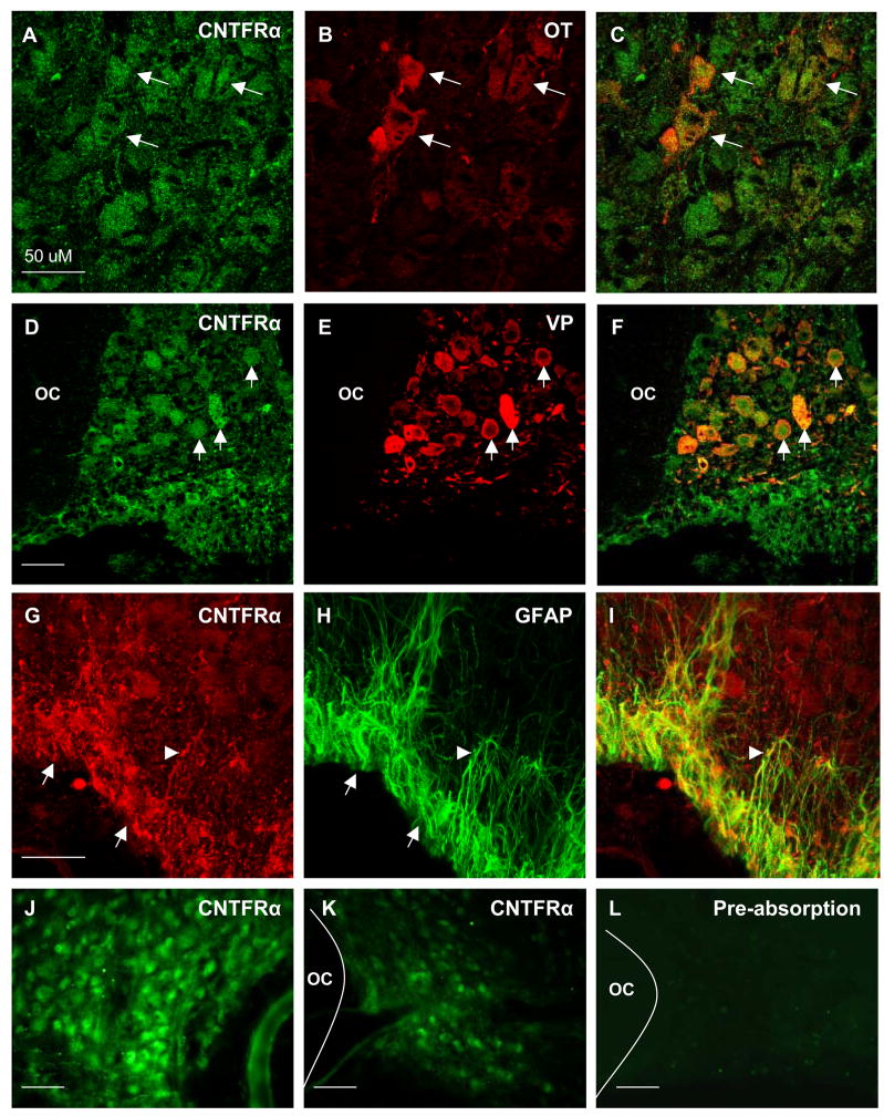
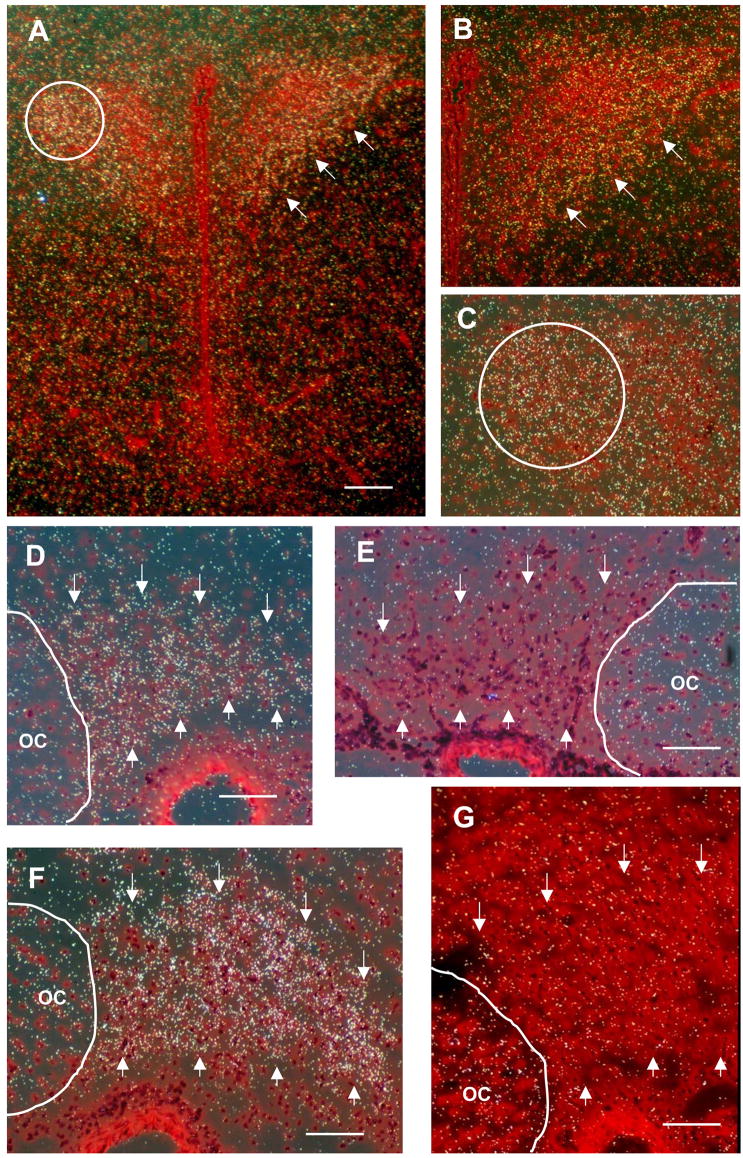
Ciliary neurotrophic factor (CNTF) is expressed by glial cells at multiple levels of the magnocellular neurosecretory system (MNS). CNTF is present in astrocytes in the hypothalamic supraoptic nucleus (SON) as well as in perivascular cells in the neurohypophysis, and a several fold increase in CNTF immunoreactivity occurs in the SON following either axotomy of magnocellular neurons or during axonal sprouting by intact magnocellular neurons. CNTF also promotes survival and stimulates process outgrowth from magnocellular neurons in vitro. While these findings suggest that CNTF may act as a growth factor in support of neuronal plasticity in the MNS, little is known regarding possible expression of receptors for CNTF in the MNS. We have therefore used immunocytochemistry and in situ hybridization to examine the expression of CNTF receptor alpha (CNTFRalpha) in the rat MNS. Robust immunoreactivity for CNTFRalpha was observed associated with oxytocinergic and vasopressinergic neurons distributed throughout the SON. Astrocytes located within the ventral glial lamina (VGL) of the SON were also immunoreactive for CNTFRalpha. Robust hybridization of an anti-sense [(35)S]-cRNA probe to CNTFRalpha mRNA was observed throughout the SON, while binding of a control sense probe was much lower. Grains were found clustered predominantly over neuronal somata, indicative of expression by magnocellular neurons within the SON. We next examined changes in expression of CNTFRalpha mRNA by magnocellular neurons 7 days following unilateral transection of the hypothalamo-neurohypophysial tract. The level of CNTFRalpha mRNA was increased 32% (compared to age-matched intact controls; p<0.05) in magnocellular neurons in the SON contralateral to the lesion, which are undergoing extensive collateral axonal sprouting, but was unchanged in axotomized magnocellular neurons in the SON ipsilateral to the lesion. These findings suggest that CNTF produced by MNS glia and acting via CNTFRalpha may exert neurotrophic effects on magnocellular neurons.
Figures
References
-
- Adams JH, Daniel PM, Prichard MMI. Degeneration and regeneration of hypothalamic nerve fibers in the neurohypophysis after pituitary stalk section in the ferret. J Comp Neurol. 1969;135:121–144. - PubMed
-
- Alonzi T, Middleton G, Wyatt S, Buchman V, Betz UAK, Muller W, Musiani P, Poli V, Davies AM. Role of STAT3 and PI 3-kinase/Akt in mediating the survival actions of cytokines on sensory neurons. Mol Cell Neurosci. 2001;18:270–282. - PubMed
-
- Antunes JL, Louis KM, Huang S, Zimmerman E. Section of the pituitary stalk in the rhesus monkey: morphological and endocrine observations. Ann Neurol. 1980;8:308–316. - PubMed
-
- Armstrong WE, Warach S, Hatton GI, McNeill TH. Subnuclei in the hypothalamic paraventricular nucleus: A cytoarchitectural, horseradish peroxidase and immunocytochemical analysis. Neuroscience. 1980;5:1931–1958. - PubMed
-
- Beck E, Daniel PM, Prichard ML. Regeneration of hypothalamic nerve fibers in the goat. Neuroendocrinology. 1969;5:161–182. - PubMed
Publication types
MeSH terms
Substances
Grants and funding
LinkOut - more resources
Full Text Sources
