. 2008 Dec 26;283(52):36354-60.
doi: 10.1074/jbc.M806143200. Epub 2008 Oct 30.

IMP dehydrogenase type 1 associates with polyribosomes translating rhodopsin mRNA

Affiliations

IMP dehydrogenase type 1 associates with polyribosomes translating rhodopsin mRNA

Sarah E Mortimer et al. J Biol Chem. .

Abstract

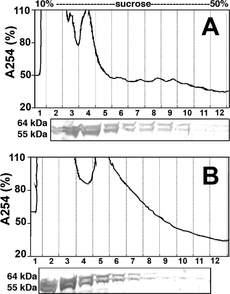
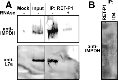
IMP dehydrogenase (IMPDH) catalyzes the pivotal step in guanine nucleotide biosynthesis. Here we show that both IMPDH type 1 (IMPDH1) and IMPDH type 2 are associated with polyribosomes, suggesting that these housekeeping proteins have an unanticipated role in translation regulation. This interaction is mediated by the subdomain, a region of disputed function that is the site of mutations that cause retinal degeneration. The retinal isoforms of IMPDH1 also associate with polyribosomes. The most common disease-causing mutation, D226N, disrupts the polyribosome association of at least one retinal IMPDH1 isoform. Finally, we find that IMPDH1 is associated with polyribosomes containing rhodopsin mRNA. Because any perturbation of rhodopsin expression can trigger apoptosis in photoreceptor cells, these observations suggest a likely pathological mechanism for IMPDH1-mediated hereditary blindness. We propose that IMPDH coordinates the translation of a set of mRNAs, perhaps by modulating localization or degradation.


Figures

FIGURE 1.
FIGURE 1.
The adRP/LCA-causing mutations of IMPDH1. A, positions of the disease-associated mutations are depicted on a monomer of IMPDH from Streptococcus pyogenes, which corresponds to the canonical IMPDH1(514) (Protein Data Bank accession number 1ZFJ (30); note that the CBS domains are disordered in the structure of human IMPDH1 (Protein Data Bank accession number 1JCN), so that several of the positions of mutation are not observed). Magenta denotes mutations that are clearly pathogenic; red, likely pathogenic; green, possibly pathogenic (27). Molecular graphics images were produced using the University of California, San Francisco, Chimera package from the Resource for Biocomputing, Visualization, and Informatics at the University of California, San Francisco (supported by NIH Grant P41 RR-01081) (14). B, scheme showing the differences between IMPDH1(514), IMPDH1(546), and IMPDH1(595).
FIGURE 2.
FIGURE 2.
Polyribosome profiles of HeLa cell extracts. Lysates were prepared and fractionated on a sucrose gradient as described under “Experimental Procedures.” A typical profile is shown, with the corresponding Western blot with anti-IMPDH antisera immediately below. For the remaining panels, fractions were obtained similarly, and Western blots were probed with antibody against the designated protein. Puromycin disrupts both polyribosomes and the high molecular weight IMPDH complex. A single blot was split and probed with anti-CBP80 antibody and anti-IMPDH antisera. CBP80 is found in the cytosolic ribosomal subunits and monosome-containing fractions, but excluded from higher density fractions, whereas IMPDH is found throughout the gradient. The bottom panel shows the sedimentation of purified recombinant (r) IMPDH1(514).
FIGURE 3.
FIGURE 3.
The association of IMPDH with polyribosomes is mediated by the subdomain. HeLa cells were transfected to express wild-type (WT) IMPDH1(514) tagged with EGFP at the C terminus (IMPDH1-GFP, WT) or IMPDH lacking subdomain (ΔSD-IMPDH1-GFP, ΔSD). Lysates were prepared, and fractions were aliquoted as described in Fig. 2 and probed with antibodies against GFP. A, IMPDH1-GFP; B, IMPDH1-GFP + puromycin; C, IMPDH1-GFP + RNase; D, GFP alone, ΔSD-IMPDH1-GFP, IMPDH1-GFP/D226N, and wild type. The IMPDH1-GFP/D226N and WT are from parallel transfections.
FIGURE 4.
FIGURE 4.
The retinal isoforms IMPDH1(546) and IMPDH1(595) associate with polyribosomes in bovine retinal cells. Lysates were prepared from freshly dissected bovine retina and fractionated as described above. Immunoblots were probed with anti-IMPDH serum BRD5. A, no pretreatment; B, RNase-treated retinal lysate.
FIGURE 5.
FIGURE 5.
The D226N mutation decreases the association of IMPDH1(595) with polyribosomes. A, lysates were prepared from HEK cells expressing GFP-tagged retinal IMPDH1 isoforms, and total polyribosomes were collected by sedimentation through a sucrose cushion. Samples were analyzed by immunoblotting with monoclonal antibody recognizing IMPDH. B, composite data from three experiments as in A. The endogenous IMPDH band was used to normalize the amount of GFP-tagged IMPDH1 in the polyribosomes. The units are arbitrary.
FIGURE 6.
FIGURE 6.
IMPDH1 is associated with polyribosomes translating rhodopsin mRNA. A, polyribosomes were isolated from bovine retinal lysate by sedimentation through a sucrose cushion as described under “Experimental Procedures.” Polyribosomes expressing rhodopsin were isolated by immunoprecipitation (IP) with monoclonal antibody RET-P1, which recognizes the N-terminal peptide of rhodopsin. The resulting precipitates were analyzed by SDS-PAGE and immunoblotting. The input sample contains both IMPDH1 and ribosomal protein L7a. Both IMPDH and L7a co-precipitate with RET-P1. Neither IMPDH1 nor L7a are observed in mock immunoprecipitations where RET-P1 is omitted or when the sample is pretreated with RNase to disrupt the polyribosomes. B, samples are prepared as in A. IMPDH1 precipitates with RET-P1 but not with monoclonal antibody 1D4, which recognizes the C-terminal segment of rhodopsin.
FIGURE 7.
FIGURE 7.
Co-immunoprecipitation of IMPDH1(546)-GFP and rhodopsin mRNA. HEK293 cells were co-transfected with constructs directing the expression of IMPDH1(546)-GFP and rhodopsin mRNA. IMPDH1(546)-GFP was immunoprecipitated with anti-GFP antibody. A, Western blot of the immunoprecipitate showing efficient pulldown of IMPDH1(546)-GFP. HEK cells transfected with constructs expressing only rhodopsin (-) and rhodopsin and IMPDH1(546)-GFP (+); ft, flow-through. B, reverse transcription (RT) followed by PCR demonstrates the presence of rhodopsin mRNA in the immunoprecipitate. Rho, rhodopsin; ft, flow-through.

References

    1. Weber, G., Nakamura, H., Natsumeda, Y., Szekeres, T., and Nagai, M. (1992) Adv. Enzyme Regul. 32 57-69 - PubMed
    1. Nimmesgern, E., Black, J., Futer, O., Fulghum, J. R., Chambers, S. P., Brummel, C. L., Raybuck, S. A., and Sintchak, M. D. (1999) Protein Expression Purif. 17 282-289 - PubMed
    1. Gan, L., Petsko, G. A., and Hedstrom, L. (2002) Biochemistry 41 13309-13317 - PubMed
    1. Janosik, M., Kery, V., Gaustadnes, M., Maclean, K. N., and Kraus, J. P. (2001) Biochemistry 40 10625-10633 - PubMed
    1. Scott, J. W., Hawley, S. A., Green, K. A., Anis, M., Stewart, G., Scullion, G. A., Norman, D. G., and Hardie, D. G. (2004) J. Clin. Investig. 113 274-284 - PMC - PubMed

Publication types

LinkOut - more resources