. 2008 Nov 1;68(21):8733-42.
doi: 10.1158/0008-5472.CAN-08-1968.

FoxM1B transcriptionally regulates vascular endothelial growth factor expression and promotes the angiogenesis and growth of glioma cells

Affiliations

FoxM1B transcriptionally regulates vascular endothelial growth factor expression and promotes the angiogenesis and growth of glioma cells

Yujian Zhang et al. Cancer Res. .

Abstract

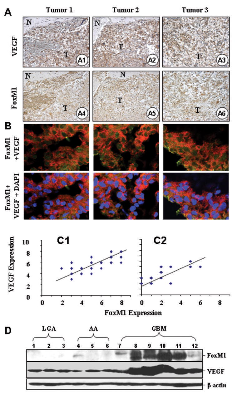
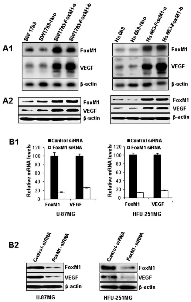
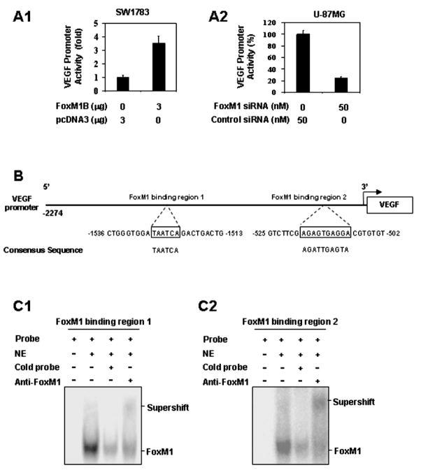
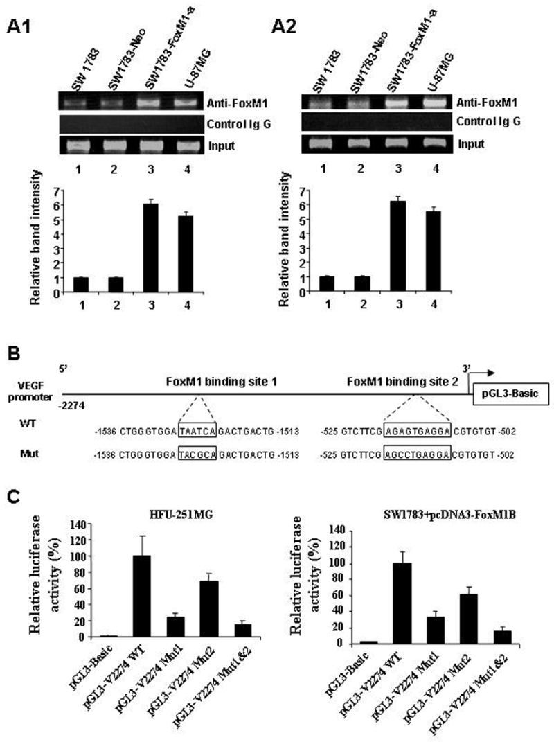
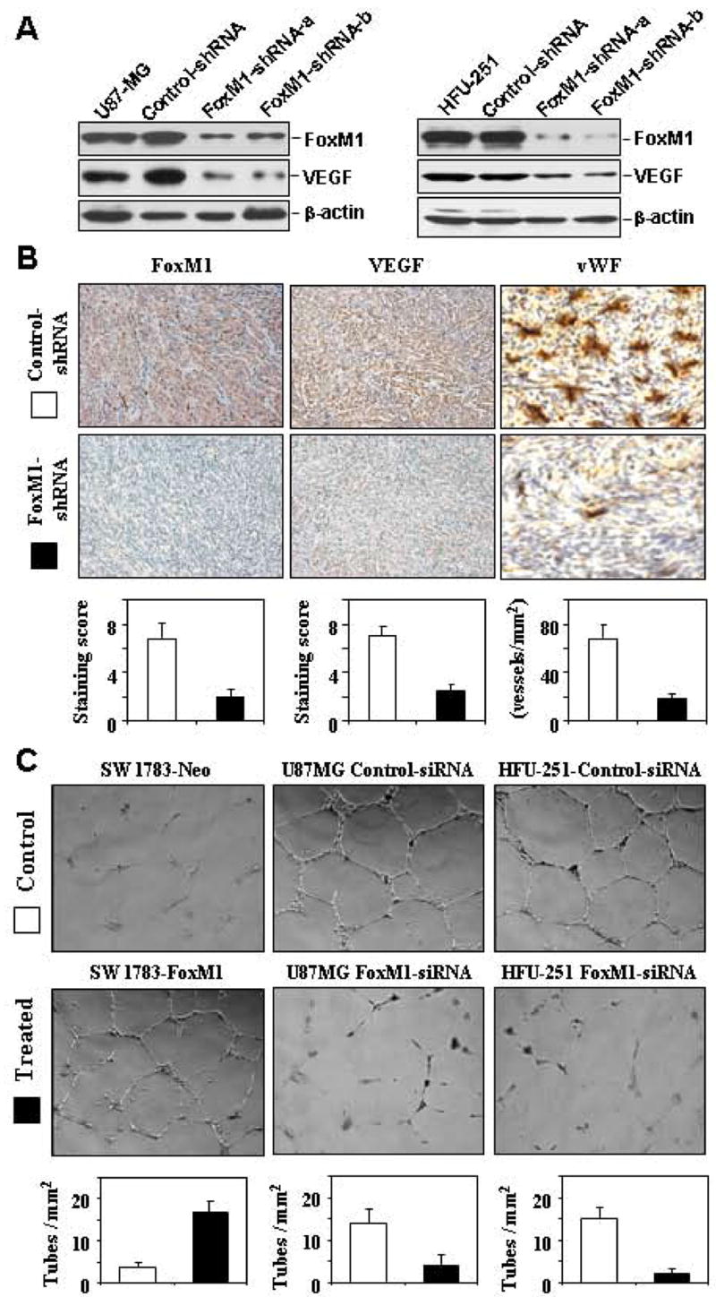
We previously found that FoxM1B is overexpressed in human glioblastomas and that forced FoxM1B expression in anaplastic astrocytoma cells leads to the formation of highly angiogenic glioblastoma in nude mice. However, the molecular mechanisms by which FoxM1B enhances glioma angiogenesis are currently unknown. In this study, we found that vascular endothelial growth factor (VEGF) is a direct transcriptional target of FoxM1B. FoxM1B overexpression increased VEGF expression, whereas blockade of FoxM1 expression suppressed VEGF expression in glioma cells. Transfection of FoxM1 into glioma cells directly activated the VEGF promoter, and inhibition of FoxM1 expression by FoxM1 siRNA suppressed VEGF promoter activation. We identified two FoxM1-binding sites in the VEGF promoter that specifically bound to the FoxM1 protein. Mutation of these FoxM1-binding sites significantly attenuated VEGF promoter activity. Furthermore, FoxM1 overexpression increased and inhibition of FoxM1 expression suppressed the angiogenic ability of glioma cells. Finally, an immunohistochemical analysis of 59 human glioblastoma specimens also showed a significant correlation between FoxM1 overexpression and elevated VEGF expression. Our findings provide both clinical and mechanistic evidence that FoxM1 contributes to glioma progression by enhancing VEGF gene transcription and thus tumor angiogenesis.


Figures

Fig. 1
Fig. 1
FoxM1 overexpression correlates with upregulation of VEGF expression in human GBM specimens. Immunohistochemical staining using specific anti-FoxM1 and anti-VEGF antibodies were performed on 59 GBM and 25 AA tissues. A, Representative photos of three tumors indicate the pattern of VEGF and FoxM1 expression in GBM sections (A1 ~ A6, “N”, normal, “T”, tumor). B, Colocalization of FoxM1 and VEGF in GBM tissues. Frozen sections of GBMs were processed for triple immunofluorescence staining for FoxM1 (green), VEGF (red) and nuclei (DAPI, blue). These representative photos of three tumors indicate that many cells displayed strong positive nuclei staining, but weak positive cytoplasma staining of FoxM1, whereas VEGF was predominantly in the cytoplasm of the cells. Merged photos indicated that FoxM1 and VEGF colocalized in the cells. C, We quantitatively scored the tissue sections according to the percentage of positive cells and staining intensity as described in “Materials and Methods”. We then combined the percentage and intensity scores to obtain a total score (range, 0 8). FoxM1 expression levels correlated positively with VEGF expression levels in GBM samples (Pearson’s correlation test r = 0.79; P < 0.001), and in AA samples (r = 0.75; P < 0.001). Note that some of the dots on the graphs represented more than one specimen (some scores overlapped). D, FoxM1 and VEGF protein expression levels from 3 LGA, 3 AA, and 6 GBM frozen tissues were determined using Western blot analysis.
Fig. 2
Fig. 2
Effects of altered FoxM1 expression on VEGF expression in human glioma cell lines. A, Upregulation of VEGF mRNA and protein expression by FoxM1B overexpression. FoxM1 and VEGF expression levels in parental, control-vector transfected, and FoxM1 expression-vector transfected SW1783 and Hs683 cells were determined by Northern blot analyses (A1), and Western blot analyses (A2). B, Downregulation of VEGF mRNA and protein expression by knockdown of FoxM1 expression. FoxM1 and VEGF expression in U-87MG and HFU-251MG cells transfected with FoxM1-siRNA or control siRNA were determined by real-time RT-PCR (B1) and Western blot analyses (B2).
Fig. 3
Fig. 3
The VEGF gene as a transcriptional target of FoxM1. A, Transactivation of the VEGF promoter by FoxM1 and repression of the VEGF promoter by FoxM1-siRNA. A1, Effects of FoxM1 overexpression on VEGF promoter activity. SW1783 and Hs683 cells were cotransfected with 1 μg of the VEGF promoter-luciferase construct pGL3-V2274 and 3 μg of pcDNA 3.1-FoxM1B or pcDNA 3.1. Activation was calculated relative to cells transfected with pcDNA 3.1. A2, Effect of FoxM1-siRNA on VEGF promoter activity. HFU-251MG and U-87MG cells were cotransfected with 1 μg pGL3-V2274 and 50 nM FoxM1-siRNA or control siRNA. Inhibition was calculated as a percentage relative to cells transfected with control siRNA. B, Sequences and positions of putative FoxM1-binding elements on the VEGF promoter. C, Binding of FoxM1 to the VEGF promoter in vitro. We performed EMSA using nuclear protein (NE) extracted from HFU-251MG cells and the oligonucleotide probes of putative FoxM1-binding region 1 (C1) and region 2 (C2) of VEGF promoter. A major shifted band was noted and was competed out by an excess of unlabeled FoxM1-binding region 1 and region 2 oligonucleotides, respectively, and supershifted by an anti-FoxM1 antibody.
Fig. 4
Fig. 4
Binding of FoxM1 to the VEGF promoter in vivo and mutational analysis of the FoxM1-binding sites. A, ChIP assays were performed with SW1783, SW1783-Neo, SW1783-FoxM1-a, and U-87MG cells. Chromatin fragments of the cells were immunoprecipitated with an anti-FoxM1 antibody (top panel) or control IgG (middle panel) and subjected to PCR. We subjected 1% of the total cell lysates to PCR before immunoprecipitation as inputs (bottom panel). The relative band intensity was calculated as the ratio between precipitated and input DNA from each cell line. Data shown are the mean values obtained from three experiments. B, Schematic structure of the VEGF promoter. The sequences of the FoxM1-binding sites are shown in both wild-type (WT) and mutant forms. C, VEGF promoter activity with and without mutations in the FoxM1-binding sites. HFU-251MG cells were transfected with the wild-type VEGF promoter or its mutants. Luciferase activity was then determined. Also, SW1783 cells were cotransfected with the wild-type VEGF promoter or its mutants and pcDNA 3.1-FoxM1 (3 g), and luciferase activity was again determined.
Fig. 5
Fig. 5
Effects of altered FoxM1 expression on VEGF expression and angiogenic potential of glioma cells. A, Western blot analyses of FoxM1 knockdown and VEGF expression in stable FoxM1-shRNA-transfected U-87MG and HFU-251MG cells (FoxM1-shRNA-a, FoxM1-shRNA-b) and controls (control-shRNA). B, Brain tumors produced by control-shRNA or FoxM1-shRNA-transfected U-87MG cells were processed and sectioned for immunostaining with specific antibodies against FoxM1 and VEGF. The tumor microvessel densities in the samples were determined by staining with an anti-factor VIII antibody. The staining patterns and quantifications shown were representative of those observed in 10 random fields. Note that knocking down FoxM1 expression inhibited VEGF protein expression and decreased microvessel densities in brain tumors. C, The angiogenic potential of glioma cells was determined by an endothelial cell tube formation assay. Samples of conditioned media were prepared from SW1783-Neo, SW1783-FoxM1, and control-shRNA or FoxM1 shRNA-transfected U-87MG or HFU-215MG cells. Human umbilical cord endothelial cells (2 × 10⁴) in 300 μL of conditioned media were then plated on growth factor reduced Matrigel to form a capillary tube. Capillary tube formation in each group was photographed and quantified. The results shown are representative of two experiments.

Similar articles

Cited by

References

    1. Kleihues P, Burger PC, Scheithauer BW. The new WHO classification of brain tumours. Brain Pathol. 1993;3:255–68. - PubMed
    1. Folkman J. Angiogenesis in cancer, vascular, rheumatoid and other disease. Nat Med. 1995;1:27–31. - PubMed
    1. Feldkamp MM, Lau N, Rak J, Kerbel RS, Guha A. Normoxic and hypoxic regulation of vascular endothelial growth factor (VEGF) by astrocytoma cells is mediated by Ras. Int J Cancer. 1999;81:118–24. - PubMed
    1. de la Iglesia N, Konopka G, Puram SV, et al. Identification of a PTEN-regulated STAT3 brain tumor suppressor pathway. Genes Dev. 2008;22:449–62. - PMC - PubMed
    1. Rahaman SO, Harbor PC, Chernova O, et al. Inhibition of constitutively active Stat3 suppresses proliferation and induces apoptosis in glioblastoma multiforme cells. Oncogene. 2002;21:8404–13. - PubMed

Publication types

MeSH terms