. 2008 Nov 1;68(21):8761-9.
doi: 10.1158/0008-5472.CAN-08-1058.

c-Cbl interacts with CD38 and promotes retinoic acid-induced differentiation and G0 arrest of human myeloblastic leukemia cells

Affiliations

c-Cbl interacts with CD38 and promotes retinoic acid-induced differentiation and G0 arrest of human myeloblastic leukemia cells

Miaoqing Shen et al. Cancer Res. .

Abstract

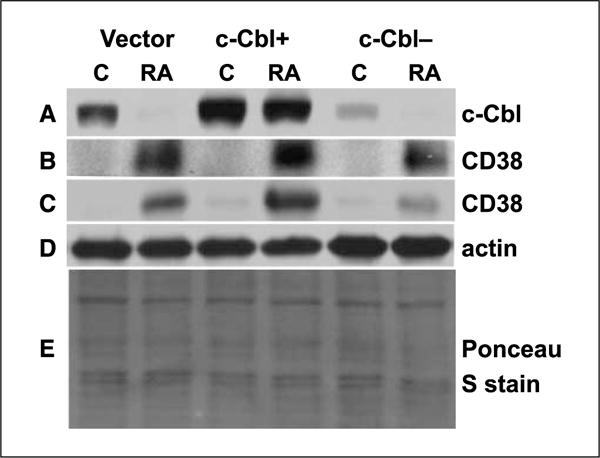
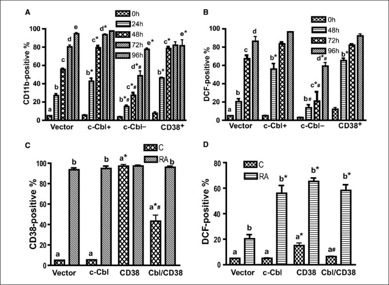
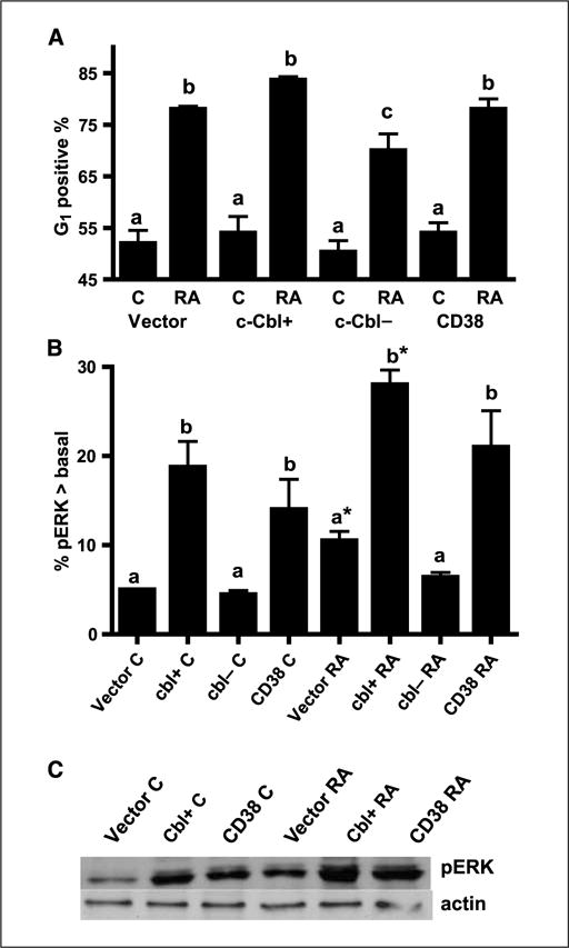
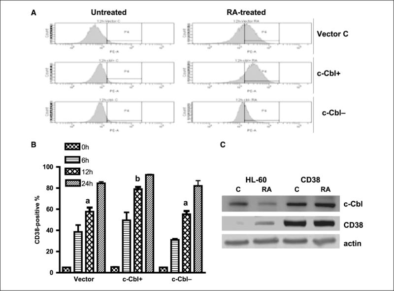
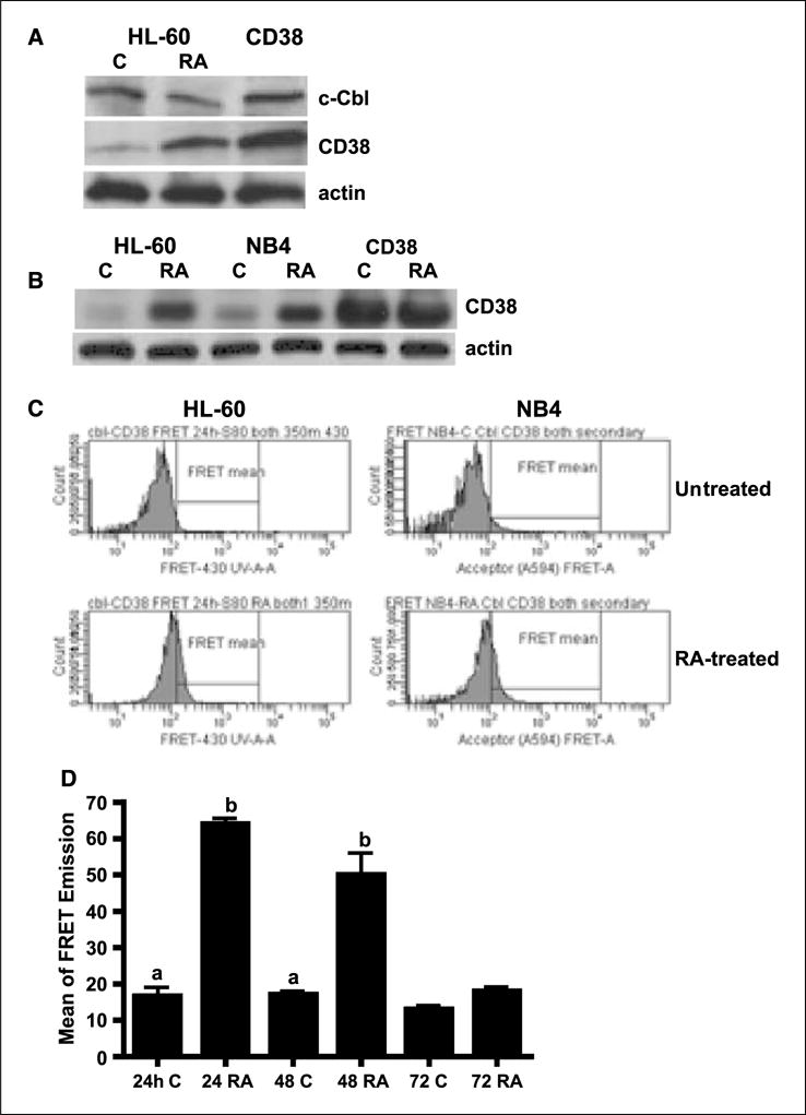
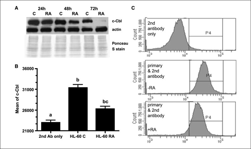
Retinoic acid (RA) is known to regulate cell growth and differentiation. In HL-60 human myeloblastic leukemia cells, it causes mitogen-activated protein kinase (MAPK) signaling leading to myeloid differentiation and G(0) cell cycle arrest. This communication reports that expression of the Cbl adaptor caused enhanced extracellular signal-regulated kinase 2 activation and promoted RA-induced differentiation and G(0)-arrest. Stable transfectants ectopically expressing c-Cbl underwent myeloid differentiation faster than wild-type (wt) cells when treated with RA. In contrast, c-Cbl knockdown stable transfectants differentiated slower than wt cells when treated with RA. Cells ectopically expressing c-Cbl had enhanced CD38 expression when treated with RA, and cells ectopically expressing CD38 had enhanced c-Cbl expression, even without with RA, suggesting an interaction between c-Cbl and CD38. Fluorescence resource energy transfer and coimmunoprecipitation showed that c-Cbl and CD38 bind each other. RA causes the gradual down-regulation and eventual loss of c-Cbl expression, resulting in loss of the Cbl-CD38 interaction, suggesting that c-Cbl plays a relatively early role in promoting RA-induced differentiation. RA-induced differentiation can thus be propelled by c-Cbl and by CD38, both of which bind together, enhance the expression of each other, and cause MAPK signaling. There thus seems to be a cooperative role for c-Cbl and CD38, reflected in their direct binding, in propulsion of RA-induced differentiation.


Conflict of interest statement

Disclosure of Potential Conflicts of Interest

No potential conflicts of interest were disclosed.

Figures

Figure 1
Figure 1
c-Cbl expression in c-Cbl stable transfectants (c-Cbl+) and knockdown (c-Cbl−) stable transfectant cell lines. Western blot of c-Cbl (72 h; A), CD38 (72 h; B), CD38 (10 h; C), and β-actin (D) expression in vector control, c-Cbl+, and c-Cbl siRNA-targeted stable transfectant cells. Protein lysates extracted from different cells were probed with anti-c-Cbl, anti-CD38, and anti-β-actin. β-Actin (D) and Ponceau S stain (E) were used to check uniform loading of protein lysates at 10 and 72 h, respectively. The blots verified c-Cbl overexpression and knockdown, and the Western blot also showed that for RA-treated cells, there was more CD38 in c-Cbl+ and less CD38 in c-Cbl knockdowns compared with vector controls.
Figure 2
Figure 2
c-Cbl, CD38, and c-Cbl/CD38 double transfectants underwent enhanced myeloid differentiation. A, the expression level of CD11b was higher in c-Cbl stable transfectant cells than that of vector control, and the loss of c-Cbl led to a decreased CD11b expression. Vector controls, c-Cbl transfectant (c-Cbl+), and siRNA targeting c-Cbl transfectant (c-Cbl−) cells were treated with RA for the indicated times and stained with APC-conjugated anti-CD11b antibody, and the percent of cells expressing CD11b was analyzed by flow cytometry. B, c-Cbl enhanced RA-induced expression of a functional differentiation marker, inducible oxidative metabolism. Vector control, c-Cbl+, and c-Cbl− cells were treated with RA for the indicated times, and the percentage of cells capable of inducible oxidation metabolism detected by DCF fluorescence was analyzed by flow cytometry. Cells were incubated in PBS containing DCF and TPA and analyzed by flow cytometry. The threshold to determine percent positive cells was set to exclude 95% of control cells. *, CD11b or DCF expression levels from c-Cbl, c-Cbl−, or CD38 were significantly different from vector controls; #, c-Cbl− transfectants were significantly (P ≤ 0.05) different from c-Cbl+ or CD38 transfectants. The different letters indicate different time points. C, percent of vector control, c-Cbl, CD38, and c-Cbl/CD38 transfectants expressing CD38 before and after 48 h of RA treatment. c-Cbl/CD38 double transfectants had less CD38 expression than wt CD38 stable transfectants. Cells were stained with PE-conjugated anti-CD38 antibody and analyzed by flow cytometry. D, compared with vector controls, c-Cbl/CD38 double transfectants had enhanced expression of a functional differentiation marker, inducible oxidative metabolism, after 48 h of RA treatment similar to c-Cbl or CD38 stable transfectant. *, significant (P ≤ 0.05) difference in same group (untreated or RA-treated); #, c-Cbl/CD38 transfectants were significantly different from CD38 transfectants. The different letters indicate significant difference between untreated and RA-treated in same cell lines.
Figure 3
Figure 3
c-Cbl accelerated G0 arrest induced by RA treatment, and both c-Cbl and CD38 enhanced ERK activation. A, percent of vector control, c-Cbl transfectants (c-Cbl+), siRNA-targeted c-Cbl transfectants (c-Cbl−), and CD38 transfectant cells with G1-G0 DNA after 72 h of RA treatment. Cells stained with hypotonic propidium iodine staining solution were analyzed by flow cytometry. The different letters indicate significant difference at the P ≤ 0.05 levels. c, for RA-treated cells, the G1 positive percentage of c-Cbl− transfectant cells is significantly (P ≤ 0.05) lower than that in vector control, c-Cbl+, and CD38 transfectants. B, percentage of cells with pERK exceeding basal levels in untreated controls. Ectopic high expression of c-Cbl and CD38 enhanced activated ERK expression and c-Cbl knockdowns did not activate ERK expression as indicated by flow cytometry. Cells were fixed, permeabilized, and stained with Alexa Fluor 647–conjugated anti-phospho-p44/42 MAPK antibody. Enhancement was measured as a positive shift of the histogram above basal levels in control cells. Induced ERK activation on RA treatment for 15 h was indicated by a shift in percentage of cells with activated ERK above the basal levels of control cells. The threshold for positive shift was set to exclude 95% of control cells with basal level pERK expression. The different letters indicate significant difference at the P ≤ 0.05 levels. *, significant (P ≤ 0.05) difference between untreated and RA-treated in same cell lines. C, Western blot of pERK for vector control, c-Cbl+, and CD38 stable transfectants (top) and β-actin (bottom). C, untreated control; RA, RA-treated for 24 h. The data from the Western blotting of pERK were consistent with flow cytometry, indicating that c-Cbl and CD38 enhanced ERK expression.
Figure 4
Figure 4
c-Cbl and CD38 enhanced the expression of each other. A, representative CD38 expression histograms of untreated and RA-treated (12 h) vector control, c-Cbl stable transfectants, and knockdown cells. B, percent of vector control, c-Cbl transfectant (c-Cbl+), and siRNA-targeted c-Cbl transfectant (c-Cbl−) cells expressing CD38 after RA treatment for the indicated times. RA-treated c-Cbl+ cells had increased expression of CD38 and c-Cbl− cells had decreased CD38 expression compared with vector control cells. The expression levels were significantly different after 12 h of RA treatment (P ≤ 0.003). Cells were stained with PE-conjugated anti-CD38 antibody and analyzed by flow cytometry. The different letters indicate significant differences between groups. C, Western blots of c-Cbl (top), CD38 (middle), and β-actin (bottom) in RA-treated (48 h) and untreated HL-60 and CD38 transfectant cells. c-Cbl expression in CD38 stable transfectant cells was enhanced. Protein lysates were extracted from wt HL-60 and CD38 overexpression cell lines and probed with anti-c-Cbl, anti-CD38, and anti-β-actin.
Figure 5
Figure 5
The interaction between c-Cbl and CD38 was confirmed by immunoprecipitation and FRET. A, Western blots of immunoprecipitated c-Cbl (top) and CD38 (middle) in HL-60 cells that were untreated (C) or treated with RA or transfected with CD38. β-Actin (bottom) was used to show input total cellular proteins in the coimmunoprecipitation reactions. Proteins immunoprecipitating with c-Cbl using anti-c-Cbl from protein lysates of wt HL-60 (untreated and RA-treated for 24 h) and CD38 stable transfectant cells were Western blotted using anti-c-Cbl (top) and anti-CD38 (middle) antibodies. Nonspecific immunoglobulin was used as a negative control. B, CD38 was induced by RA in NB4 cells and the inducible CD38 levels were the same compared with HL-60 cells. C, the interaction of c-Cbl and CD38 was observed in both HL-60 and NB4 cells by using FRET techniques. Histograms of FRET signals by flow cytometry showing the difference between 24 h of untreated and RA-treated in HL-60 and NB4 cells. D, fluorescence energy transfer from c-Cbl to CD38 for control and RA-treated HL-60 cells. There was a significant difference in energy transfer between untreated and RA-treated at 24 h (P < 0.0001) and 48 h (P = 0.03), but no difference at 72 h, suggesting an interaction between CD38 and c-Cbl after 24 and 48 h of RA-treatments. The data were analyzed by ANOVA (StatView). The different letters indicate significant difference from control cells. Cells fixed in paraformaldehyde and permeabilized were incubated in PBS containing rabbit anti-c-Cbl and mouse anti-CD38 primary antibodies and then stained with Alexa Fluor 350–conjugated and Alexa Fluor 430–conjugated goat anti-rabbit and goat anti-mouse secondary antibodies.
Figure 6
Figure 6
RA reduced c-Cbl expression in HL-60 cell line. A, Western blot of c-Cbl expression in HL-60 cells cultured without (C) or with RA for 24, 48, and 72 h showed that RA down-regulated c-Cbl expression over time. Ponceau S staining and β-actin antibody were used to check uniform loading. B, flow cytometric analysis of c-Cbl expression in untreated (C) and RA-treated (48 h) HL-60 cells. Flow cytometry data were consistent with Western blots, indicating that RA reduced c-Cbl expression. After the cells were fixed and permeabilized, they were stained with a polyclonal primary antibody against c-Cbl and a secondary Alexa Fluor 350–conjugated goat anti-rabbit antibody. Results are given as the mean fluorescence intensity from the entire gated cell population. The different letters indicate significance at the P ≤ 0.05 level. C, representative flow cytometric histograms of c-Cbl expression for untreated (middle) and RA-treated (48 h; bottom) HL-60 cells. Top, secondary antibody only staining shows background signal from the secondary antibody.

Similar articles

Cited by

References

    1. Sklan D. Vitamin A in human nutrition. Prog Food Nut Sci. 1987;11:39–55. - PubMed
    1. Mann G, Reinhardt D, Ritter J, et al. Treatment with all-trans retinoic acid in acute promyelocytic leukemia reduces early death in children. Ann Hematol. 2001;80:417–22. - PubMed
    1. Collins SJ, Gallo RC, Gallagher RE. Continuous growth and differentiation of human myeloid leukaemic cells in suspension culture. Nature. 1977;270:347–9. - PubMed
    1. Bretiman TR, Selonick SE, Collins SJ. Induction of differentiation of the human promyelocytic leukemia cell line (HL-60) by retinoic acid. Proc Natl Acad Sci U S A. 1980;77:2926–40. - PMC - PubMed
    1. Yen A. HL-60 cells as a model of growth control and differentiation: the significance of variant cells. Hematol Rev. 1990;4:5–46.

Publication types